UNIT – 9
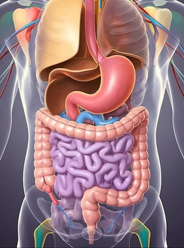
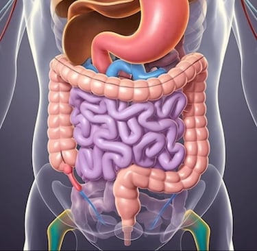
DIGESTIVE SYSTEM
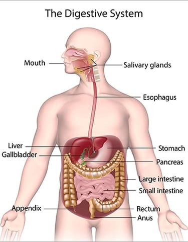
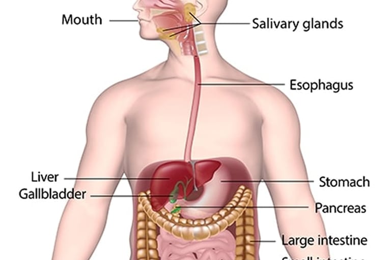
Digestive system is responsible for breaking down the food into nutrients which the body uses for energy, growth, cell repair.
It consists of series of organs and glands that work together to convert food into usable form.
ORGANS OF THE DIGESTIVE SYSTEM :
ALIMENTARY TRACT :
· It is a long hollow tube.
· Food passes through this tube.
· It start from mouth and ends in Anus.
The tube is divided into different parts,, these parts are:
(1) Mouth
(2) Pharynx
(3) Oesophagus
(4) Stomach
(5) Small intestine
(6) Large Intestine
(7) Rectum And Anal canal.
ACCESSORY ORGANS :
· These are glands in the accessory organs.
· These are second group of organs, they play role in digestion process by producing substance that help in digestion.
ACCESSORY ORGANS INCLUDE :
(1) TEETH
(2) TONGUE
(3) 3 PAIRS OF SALIVARY GLANDS
(4) LIVER AND BILIARY DUCT
(5) GALL BLADDER
(6) PANCREAS
STRUCTURE OF ALIMENTARY CANAL :
It consists of four layers.
These Are :
(A)MUCOSA :
· It is the innermost layer of the Alimentary Tract.
· It consists of three layers.
(1)MUCOUS MEMBRANE :
· It is the innermost lining of the Alimentary tract.
· It is in direct contact with the food.
· This layer is lined with Columnar epithelium.
It serves three functions :
· Protection
· Secretion
· Absorption.
It consists of Non Keratinized, stratified Squamous epithelium.
Epithelium cells Secrete mucus.
· This mucus act as a lubricant.
· When food enter the canal it prevents friction.
· There are specialized cells which Secrete their secretions into the lumen of the tract.
· The secretions include gastric juice from the gastric gland.
· Saliva from Salivary glands.
· Pancreatic juice from the pancreas.
· Bile juice from the liver.
· Intestinal juice from the intestinal glands.
(2)LAMINA PROPRIA :
· Lamina Propria is a layer of connective tissue that lies just beneath the Epithelial layer of Mucous membrane in various organs of the body.
· Lamina propria contains blood vessels.
· It consists of lymphoid tissue.
· It has a protective function.
(3)MUSCULARIS MUCOSA :
· It is a thin layer of smooth muscles.
· It provides Involution of the Mucosa layer. Example Gastric Glands, villi.
(B)SUBMUCOSA :
· It consists of areolar connective tissue.
· It is a highly vascular layer.
· It contains blood vessels and nerves.
· Submucosa contain a portion of submucosal plexus.
· The plexus regulate the movements of Mucosa.
· It also help in Vaso-constriction.
(C)MUSCULARIS (MUSCLE LAYER ) :
· Muscle layer consists of two layers of smooth muscles.
· Outer layer consists of longitudinal muscles.
· Inner layer consist of circular muscles.
· These are involuntary muscles.
· The contraction of these muscles help in breakdown of food.
· It contain the major Nerve supply myenteric plexus.
· This plexus controls the contraction of the muscularis.
· These contraction help in mixing of food.
· This type of contraction of muscles is known as peristalsis.
(D)ADVENTITIA (OUTERCOVERING) :
· It is the superficial layer.
· It consists of loose fibrous tissue
· It cover certain part of the digestive system specifically those not covered by peritoneum.
FOUND IN :
Found in retroperitoneal area or outside the peritoneal cavity.
Such as :
· Oesophagus
· Rectum
· Parts of Duodenum
PERITONEUM :
· Peritoneum is a layer that surrounds the abdominal organs including stomach and is separate from the stomach four Wall layers.
· Peritoneum is a thin serious membrane that lines the abdominal cavity and covers most of the abdominal organs.
It is made up of two layers.
(1)PARIETAL PERITONEUM :
It lines the inner surface of the abdominal wall.
(2)VISCERAL PERITONEUM :
· It covers the outer surface of the abdominal organs.
· Between these two layers is the peritoneal cavity which contains a small amount of lubricating fluid to reduce friction during organ movement.
FUNCTION OF PERITONEUM :
· It provides a sleepy surface for organs to move easily.
· It support blood vessels ,nerve vessels and lymphatic vessels Supplying the abdominal cavity.
· Play an important role in immunity and fat Storage.
NERVE SUPPLY :
The autonomic nervous system i.e Sympathetic and Parasympathetic supplies the Alimentary tract.
The Sympathetic Supply :
· These form the plexus in the Thorax ,abdomen and pelvis.
· These nerves pass to the organs of the Alimentary tract.
· Reduce the smooth muscles contraction.
· It also reduce glandular Secretion.
PARA SYMPATHETIC SUPPLY :
· The pair of Cranial nerve and Vagus nerve Supply to the Alimentary tract.
· It causes smooth muscle contraction and the secretion of digestive juice.
BLOOD SUPPLY :
ARTERIAL SUPPLY IN THE THORAX :
· The Oesophagus arteries Supply blood to the oesophagus.
· These are the branches Arise from the Thoracic Aorta.
ABDOMINAL AND PELVIC BLOOD SUPPLY :
The stomach, pancreas, spleen, liver, gallbladder and bile duct Are Supplied by Coeliac Arteries.
These are :
· Left Gastric Artery.
· Hepatic Artery.
· Splenic Artery.
The small intestine, Caecum , Ascending and Transverse colon are Supplied by Superior Mesenteric Artery.
The descending Colon, Sigmoid Colon and Rectum are Supplied by Inferior Mesenteric Artery.
VENOUS DRAINAGE :
IN THE THORAX :
· Oesophageal veins drain into azygos and hemizygous veins.
· The Azygos veins join Superior Vena Cava and Hemizygous veins join left Brachiocephalic vein.
IN THE ABDOMEN AND PELVIS :
· Portal vein is formed that drains the blood from lower part of the oesophagus, stomach, pancreas ,small intestine, large intestine and most of the Rectum.
· Portal Vein passes to the liver and then join inferior vena cava.
· Internal Iliac veins, drain blood from the lower part of the rectum and Anus.
· By passing from Hepatic Circulation the internal Iliac veins join the Inferior Vena Cava.
ALIMENTARY TRACT ORGAN :
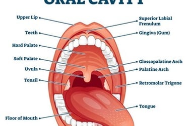
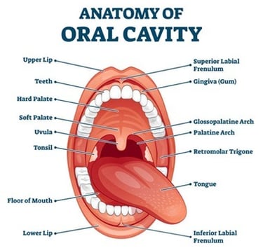
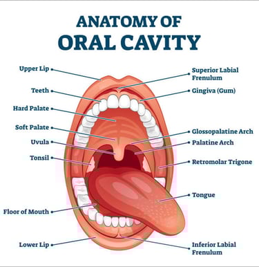
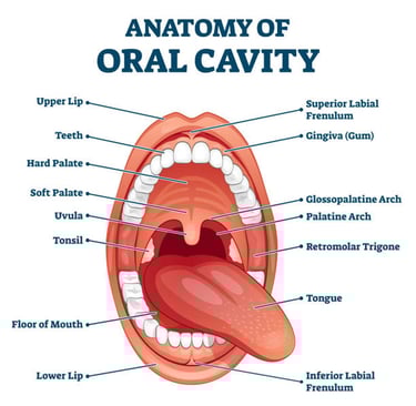
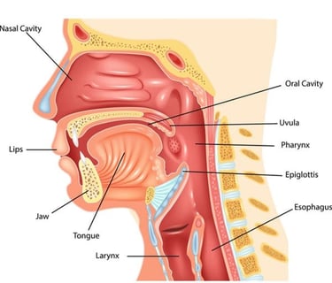
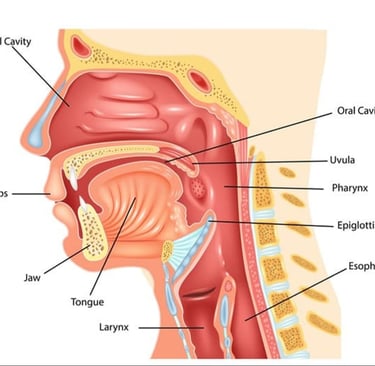
(1)MOUTH :
Mouth is also called oral cavity.
Mouth consists of muscles and bones.
ANTERIOR : LIPS
POSTERIOR : OROPHARYNX
LATERAL : MUSCLES OF CHEEK.
SUPERIOR : HARD PALATE
INFERIOR : TONGUE AND ITS MUSCLES.
EXTERNAL STRUCTURE :
(1)LIPS :
· Lips are fleshy fold surrounding the opening of the mouth.
· Inner surface of each lip is attached to the gum by a midline fold of mucus membrane called the labial frenulum.
· The Orbicularis Oris muscle and buccinator muscles in the lip help to keep food between upper and lower teeth.
(2)ORAL VESTIBULE :
The space between the gums and cheeks is called vestibule.
INTERNAL STRUCTURE :
(1)TEETH :
These are 32 in adults, used in Chewing food.
(2)GUMS :
Soft tissue surrounding and Supporting the teeth.
(3)HARD PALATE :
The Bony front part of the roof of the mouth, hard Palate is formed by Maxilla and Palatine bones.
(4)SOFT PALATE :
The soft back portion of the roof of the mouth helps with Swallowing and speech.
(5)UVULVA :
Small fleshy extension at the back of the soft palate.
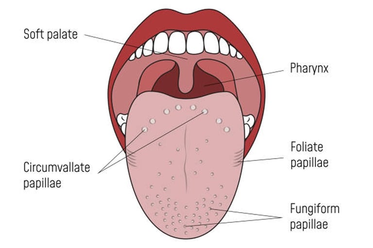
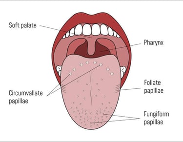
(6)TONGUE :
Muscular organ for tasting, swallowing and speaking.
(7)SALIVARY GLANDS :
They Produce Saliva to help in digestion and maintaining oral hygiene.
(2)TONGUE :
· It forms the floor of the oral cavity.
· Tongue is divided into halves by medial septum.
· It is attached inferiorly to the hydroid bone.
· The superior surface of tongue consists of stratified squamous epithelium.
· Upper and lateral surface of tongue is covered with papillae.
· The nerve Endings of sense of taste are present in it, which are also called taste buds.
There are 3 types of Papillae :
(1)FILLIFORM PAPILLAE :
These are conical projections present anteriorly over two thirds of the tongue ,they contain no taste buds.
(2)FUNGIFORM PAPILLAE :
· These are present on tip and sides.
· They appear as Red dots.
· Most of them contain taste buds.
(3)VALLATE PAPILLAE :
· These are arranged in the form of inverted V on the posterior surface of the tongue.
· These are the largest glands.
BLOOD SUPPLY TO TONGUE :
· Lingual branch of external carotid artery supplies blood to the tongue.
· Lingual veins drain the blood into internal jugular vein.
NERVE SUPPLY :
· Lingual branch of Mandibular nerves are somatic nerves which supply the area.
· Facial and Glossopharyngeal nerve, are the nerves of the special sense of taste supply the tongue area.
· Hypoglossal 12th cranial nerve supply the voluntary muscle tissue.
FUNCTION OF TONGUE :
(1)SPEECH :
The tongue helps in speech.
It manipulates the speech as we have seen in larynx.
(2)TASTE :
The taste buds are present on the surface of the tongue.
Taste buds can judge any type of taste i.e Sour, sweet, salty.
(3)MASTICATION :
It means chewing the tongue along with teeth help in chewing.
It helps in movement of food in mouth.
(4)DEGLUTITION :
It means swallowing.
The tongue pushes the food back and helps in swallowing of food.
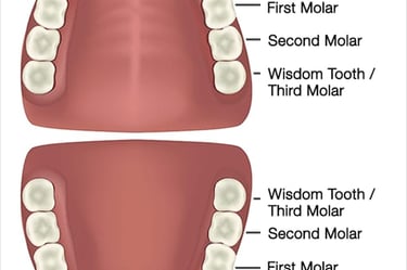
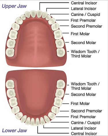
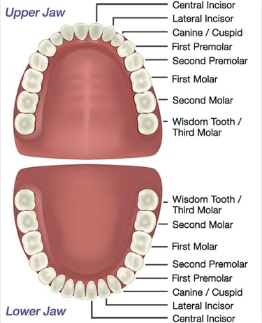
(3)TEETH :
· Teeth are fixed in jaws.
· The teeth are replaced only once.
· The teeth of first set are called milk or deciduous teeth.
· The second set are known as permanent teeth.
· The deciduous teeth are 20 in number.
· In each half of the jaw, there are 2 incisors,1 canine and 2 molars.
· The permanent teeth are 32 in number, and consists of half of each jaw 2 incisors,1 canine,2 premolars and 3 molars.
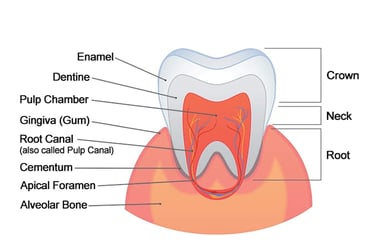
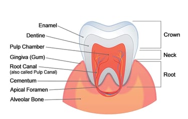
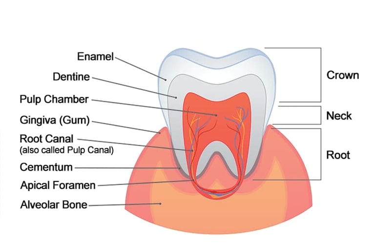
STRUCTURE OF TOOTH :
(A)ENAMEL :
Hard outer layer- the strongest substance in body.
(B)DENTIN :
Under the Enamel, less hard carries sensations.
(C)PULP :
Soft inner part with nerves and blood vessels.
FUNCTION OF TOOTH :
· Incisor and canine act as a blade, used for tearing and cutting.
· Molar and premolar are used for grinding.
BLOOD SUPPLY :
· Branches of maxillary artery supply blood to tooth.
· Venous drainage is by internal jugular veins.
NERVE SUPPLY :
· Upper teeth are supplied by branches of maxillary nerves.
· Lower teeth are supplied by branches of mandibular nerves.
· Both are branches of 5th cranial nerve, trigeminal nerve.
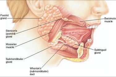
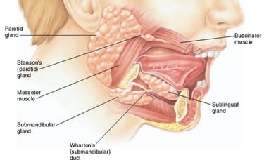
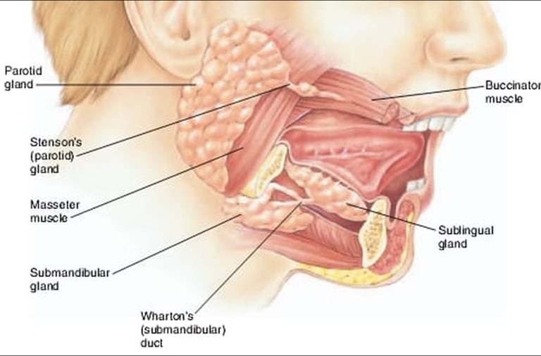
(4)SALIVARY GLANDS :
· Salivary gland are accessory organs.
· These produce a secretion called saliva.
· There are three pairs of glands.
· They help in digestion and maintain oral health.
(1)PAROTID GLANDS :
· These are located in front of the Ear below the jaw.
· These are present in pairs.
· Each gland has a parotid duct.
· This duct opens into mouth at the level of the second upper molar tooth.
· These glands produce watery saliva containing enzyme.
(2)SUB MANDIBULAR GLANDS :
· These are located between the mandibular.
· These glands produce both mucus and watery enzyme.
· The Submandibular duct open into mouth.
· Either side of the Lingual frenulum.
· These are present in pair.
(3)SUBLINGUAL GLAND :
· These are the smallest gland.
· These are also present in pair.
· The sublingual ducts open into the floor of the mouth.
STRUCTURE OF SALIVARY GLANDS :
· Each Gland is surrounded by a fibrous capsule.
· They consists of lobules.
· Lobules contain secretary cells.
· The secretions are poured into the ductules which joined to form larger ducts leading into the mouth.
NERVE SUPPLY :
· Glands are supplied by para sympathetic And sympathetic nerve fibres.
· Parasympathetic impulses in the facial and glossopharyngeal nerve stimulates the secretion of Saliva.
· Sympathetic nerve decrease the release of Saliva.
BLOOD SUPPLY :
· External Carotid artery supply the arterial blood.
· Venous drainage is into the external jugular vein.
SALIVA :
· Saliva is a clear watery fluid secreted by the Salivary glands into the mouth.
· A person produce 1 to 1.5 litre of Saliva daily.
PH of Saliva- 6.35 -6.85
COMPOSITION OF SALIVA :
· Saliva is 99% Water.
· Remaining 1% is :
· Salivary Amylase- Enzyme
· Mucus
· Electrolytes (Na+, K+,Cl-,HCO3-)
· Lysozyme
· Immunoglobulins (IgA )
· Urea and Uric acid
· Blood clotting factors.
FUNCTION OF SALIVA :
(1)Lubrication of Food :
· Saliva moistens and lubricate the food.
· It also Keep the mucus lining of the mouth intact.
(2)ORAL HYGIENE :
Saliva flushes away food particles and bacteria and helps in maintaining oral health.
(3)NON SPECIFIC DEFENSE :
Lysozyme, Clotting factors and immunoglobulins provide defence I.e they kills the harmful microorganisms and prevent from infection.
(4)TASTE :
Water present in Saliva dissolves food molecules and help the taste receptors, so they can be tasted.
(5)DIGESTION :
· It breaks down the food particles.
· It help in chemical digestion of food.
· Salivary amylase helps in partial digestion of food.
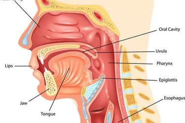
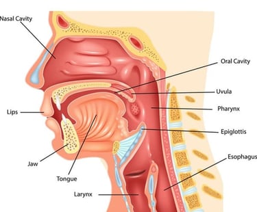
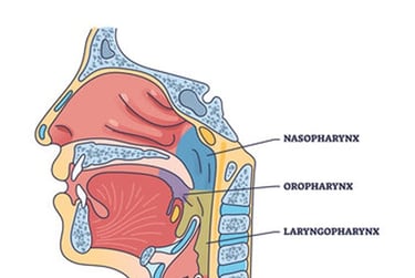
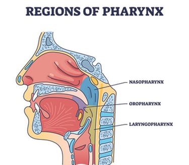
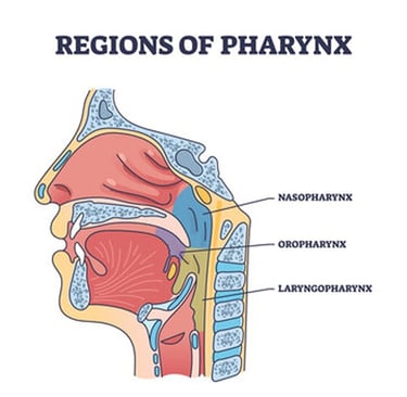
PHARYNX :
· Pharynx is a muscular tube that connects the nasal cavity and mouth to the oesophagus and larynx.
· It play role in digestion, respiration, and speech .
Pharynx is divided into:
(1)NASOPHARYNX :
Location : Behind the nasal cavity.
FUNCTION :
Air passage connect to the Eustachian tube in ear .
It is concerned with respiration.
(2)OROPHARYNX :
Situated behind the oral cavity.
FUNCTION :
Passageway for air food and liquid .
It is concerned with digestive system.
(3)LARYNGOPHARYNX :
Situated behind the larynx.
FUNCTION :
· Direct food to oesophagus .
· The food passes From Mouth to oesophagus through Laryngopharynx.
WALLS OF PHARYNX :
(1)MUCOUS LAYER :
· It is the innermost layer.
· It consists of stratified squamous epithelium.
(2)SUBMUCOUS LAYER :
· It is the middle layer.
· It consists of fibrous tissue.
· it contains blood, lymph vessels and nerves.
(3)MUSCULAR LAYER :
· It is the outer layer it consists of three muscles which help in Constriction and swallowing.
· The Passage of food through Pharynx is involuntary.
BLOOD SUPPLY :
External carotid artery and facial artery supply blood to the pharynx.
NERVE SUPPLY :
· Pharynx is supplied by Pharyngeal plexus of nerves.
· This Plexus is formed by Vagus and glossopharyngeal nerve.
· It provides parasympathetic nerve Supply.
· Sympathetic supply is from cervical ganglia.
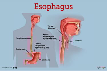
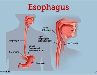
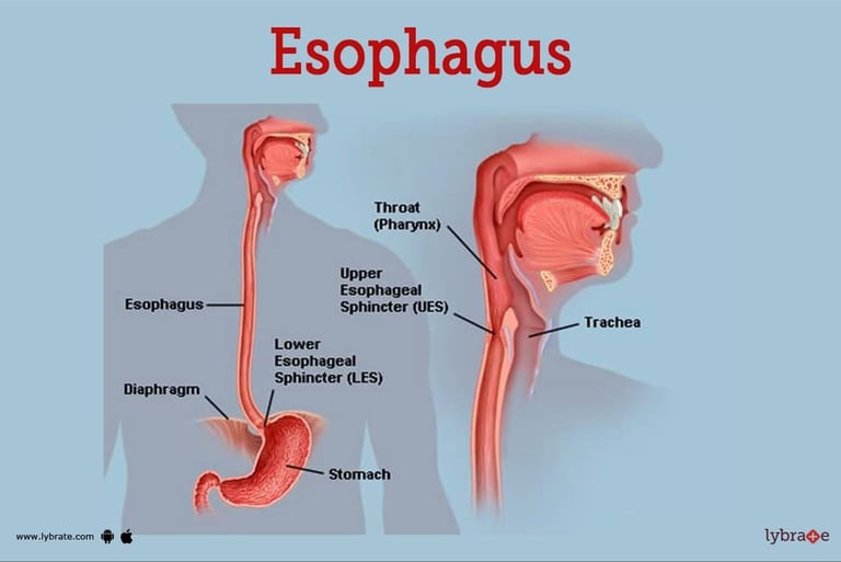
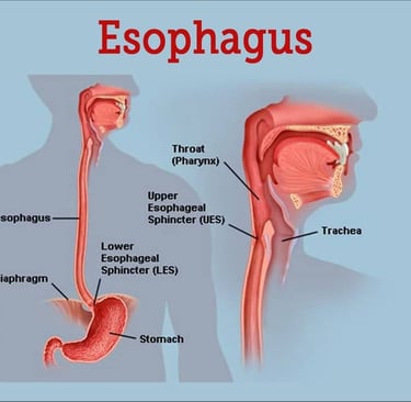
OESOPHAGUS :
Oesophagus is a wide muscular tube about 25cm long that carries food and liquid from the Pharynx to the stomach.
LOCATION :
Begins at C6.
Ends at T10 – T11
It passes through the neck , thorax and diaphragm.
OESOPHAGEAL SPHINCTER :
UPPER OESOPHAGEAL SPHINCTER :
· It is also known as Cricopharyngeal sphincter.
· It prevents air from entering the oesophagus during breathing.
LOWER OESOPHAGEAL SPHINCTER :
· It is also known as cardiac Sphincter, it prevents the reflex of acid from the stomach into the oesophagus.
ASSOCIATED STRUCTURE OF OESOPHAGUS :
ANTERIOR : Trachea
POSTERIOR : Vertebral column
Lateral : Right and Left Lung
STRUCTURE :
It consists of four layers.
(1)MUCOSA :
It is the innermost lining, it protects against abrasion.
(2)SUBMUCOSA :
It contain blood vessels and nerve endings.
(3)MUSCULARIS LAYER :
It helps in peristalsis movement in the oesophagus.
(4)ADVENTITIA :
It is the outer layer of connective tissue.
BLOOD SUPPLY :
It is supplied by thyroid and oesophageal arteries.
VENOUS DRAINAGE :
Blood from oesophagus drain into the brachiocephalic vein, from there it goes into the Azygous vein and lower end goes into the left gastric vein.
NERVE SUPPLY :
Oesophagus is supplied by both parasympathetic and sympathetic nerve fibre.
FUNCTION OF OESOPHAGUS :
(1)TRANSPORTATION :
It transport food from the mouth to stomach via Peristalsis movement.
(2)PREVENT REGURGITATION :
It prevents the backflow of the food using lower Oesophageal Sphincter.
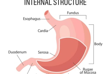
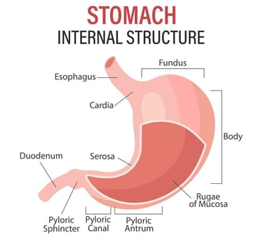
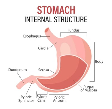
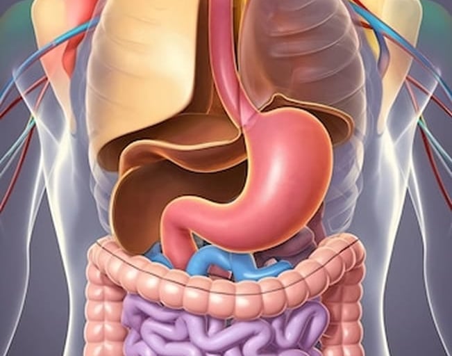
STOMACH :
· It lies in the Epigastric region of the abdomen and partially lies in the left hypochondriac region.
· It is J shaped, dilated portion of the Alimentary canal.
ASSOCIATED STRUCTURE :
ANTERIOR : Left lobe of liver.
POSTERIOR : Abdominal Aorta, pancreas, spleen , left kidney.
SUPERIOR : Diaphragm, Oesophagus.
INFERIOR : Transverse colon and small intestine.
LATERAL : LIVER AND DUODENUM
STRUCTURE OF STOMACH :
(1)FUNDUS :
· It is the upper convex Dome shape part of stomach.
· It is situated above at the level of Cardiac orifice.
· It mainly act as a storage area for undigested food and gases released during the process of digestion.
(2)BODY :
· Body of stomach is the central largest portion of the stomach located between the fundus and the pyloric part.
· It play main role in chemical digestion.
· It contains parietal cells and chief cells.
· Parietal cells secrete Hydrochloric acid and chief cell secrete pepsinogen.
· Inner surface has folds that allows expansion after meal.
(3)PYLORUS :
· Pylorus is the distal part of the stomach that connects to the duodenum.
· It plays an important role in regulating the passage of partially digested food into the intestine.
It has 2 parts :
(A)PYLORIC ANTRUM :
The wider proximal portion that receive food from the body of the stomach.
(B)PYLORIC CANAL :
The narrower part that leads to the pyloric sphincter.
FUNCTION OF PYLORUS :
· It mixes stomach content .
· Regulate the rate of gastric emptying.
The opening of stomach is guarded by two sphincter.
(1)CARDIAC SPHINCTER :
It is the point where oesophagus opens into the stomach.
(2)PYLORIC SPHINCTER :
The Pylorus connects with the duodenum of the small intestine via a sphincter called pyloric Sphincter.
CURVATURE OF STOMACH :
Stomach has 2 curvature.
(A)A Lesser Curvature is a concave and forms the right border of the stomach.
(B) The Greater Curvature is convex and forms left border of the stomach.
WALLS OF STOMACH :
The Walls of stomach consists of several layer, Each with a specific function related to digestion, protection, and movement.
The stomach has four layers.
(1)MUCOSA :
· It is the innermost Layer, that lines the stomach.
· It contains columnar epithelium tissue.
It consists of further 2 layers.
(A)LAMINA PROPRIA :
Connective tissue is present along with immune cells and blood vessels.
(B)MUSCULARIS MUCOSA :
Thin muscle layer that helps in secretion and movement.
(2)SUBMUCOSA :
· It Supports the Mucosa and provides elasticity.
· Loose connective tissue is present.
· Blood vessels, lymphatic vessels and nerves are present.
(3)MUSCULAR COAT :
It has three muscle layers.
(A)INNER OBLIQUE :
It helps in mixing of food.
(B)MIDDLE CIRCULAR LAYER :
· It forms the pyloric Sphincter.
· It helps in gastric mixing and gastric emptying.
(4)SEROSA :
· It is the outermost layer.
· A thin layer of connective tissue covered with simple Squamous epithelium.
· It reduces friction with other abdominal organs.
BLOOD SUPPLY :
· Arterial blood is Supplied by Coeliac and splenic artery.
· Stomach veins drain into Portal superior mesenteric vein and splenic veins.
NERVE SUPPLY :
· The sympathetic nerves are derived from Coeliac and Hepatic plexus.
· The parasympathetic nerves supply is from Vagus nerve.
· The parasympathetic nerve stimulation causes increased motility of stomach and secretion of gastric juice.
FUNCTION OF STOMACH :
(1)Storage of food :
It serves as a Reservoir, holds ingested food and release it gradually into the small intestine.
(2)MECHANICAL DIGESTION :
Contraction of muscular coat churn and mix Food with gastric secretions to form Chyme.
(3)CHEMICAL DIGESTION :
HCL :
Hydrochloric acid contain Acid which aid in digestion of Food and kills microorganisms.
PEPSINOGEN :
It is secreted by chief Cells of the Stomach.
Pepsinogen convert into pepsin which help in digestion of protein.
(4)SECRETION OF GASTRIC JUICE :
GASTRIN HORMONE :
· Gastrin hormone is released directly into the bloodstream.
· It Stimulate the gastric gland to produce more gastric juice.
· Gastric secretion is suppressed When the PH in the Pyloric Antrum falls to about 1.5
· The stomach Secrete about one 1.5 to 2.5 litre of gastric juice daily in the form of hydrochloric acid.
GASTRIC JUICE :
· This is a colourless fluid .
· The food gets mixed with gastric juice.
· Gastric juice is secreted about 2 litres per day.
COMPOSITION OF GASTRIC JUICE :
· Water
· Mineral Salt
· Mucus secreted by Goblet cells
· Hydrochloric acid
· Intrinsic factor
· Pepsinogen secreted by Chief Cells in the stomach gland.
FUNCTION OF GASTRIC JUICE :
· Water liquify the food.
· Hydrochloric acid acidifies All foods.
· It act as an antiseptic and kills the microorganisms taken along with food.
· Pepsin, the digestive enzyme which is obtained from Pepsinogen in presence of hydrochloric acid,, Helps in digestion of protein.
· Gastric lipase is present in the stomach, act as the digestion of fat.
(5)SECRETION OF MUCUS :
It protect the inner lining of the stomach from acid.
The Mucus act as a lubricant it prevents friction between food and internal layer of stomach, when food enters into the stomach.
(6)SECRETION OF INTRINSIC FACTOR :
Intrinsic Factor is needed for the vitamin B-12 absorption in the Ileum.
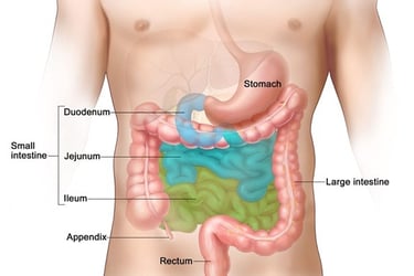
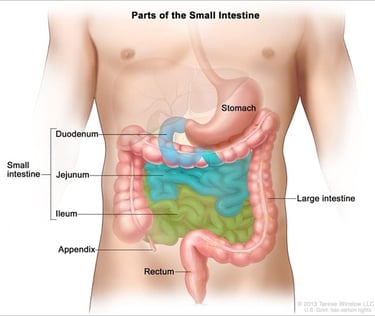
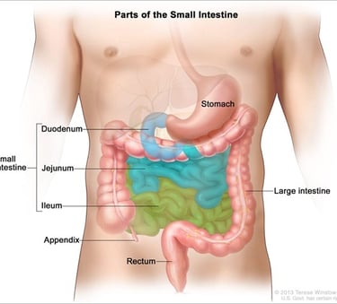
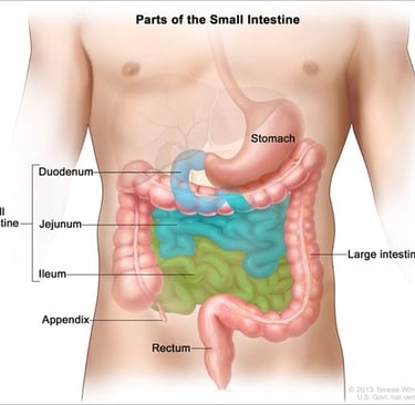
SMALL INTESTINE :
· Small intestine is a long coiled tube in digestive system.
· It is about 6 meter in length in adults.
· It extends from the stomach to the ileocecal valve where it joins large intestine.
Location : In the Umbilicus
Small intestine is divided into three parts :
(1)DUODENUM :
It is the uppermost part of small intestine.
It is about 25cm long.
It is attached to the pylorus of the stomach.
It is the first portion where partially digested food from stomach mix with bile and pancreatic juice to continue digestion.
(2)JEJUNUM :
It is about 2.5 meter in length.
It is a middle part where most nutrients are Absorbed into bloodstream.
(3)ILLEUM :
It is the third part of small intestine.
It is about 3.5 meter in length.
It joins the large intestine at the ileocecal sphincter.
It absorbs vitamin B-12, bile salt and remaining nutrients.
STRUCTURE OF SMALL INTESTINE :
It is composed of four coats.
(A)MUCOSA :
· Lined with Villi and Micro villi.
· Micro Villi increases surface area for absorption.
· It contains goblet cells, which produce mucus.
· It also contain enterocytes which help in absorption of nutrients.
· It has intestinal glands that secrete digestive enzymes.
(B)SUBMUCOSA :
· it contain Bruner’s glands, that secrete alkaline mucus.
· It contain blood vessels, lymph vessels and nerves.
(C)MUSCULAR COAT :
· The muscular coat consist of two layers :
· Outer longitudinal layer and inner circular layer.
· It is responsible for peristalsis.
(D)SEROSA :
· it is the outer covering of connective tissue covered by a layer of mesothelium.
· It is the extension of peritoneum.
PERITONEUM :
· Jejunum and ileum are completely surrounded by the peritoneum.
· It is double layer called mesentery which contain blood vessels, nerve and lymphatics.
BLOOD SUPPLY :
· Small intestine is Supplied by Superior Mesenteric Artery.
· Superior Mesenteric vein drain the blood.
NERVE SUPPLY :
Both sympathetic and parasympathetic nerve from Vagus reach the duodenum and other parts.
FUNCTION OF SMALL INTESTINE :
(1)DIGESTION :
When Chyme enters the small intestine, it gets mixed with pancreatic juice, intestinal juice and Bile juice.
(1)PANCREATIC JUICE :
It contain 3 digestive enzymes.
It is a alkaline fluid consist of :
· Water
· Mineral salt
· Enzyme
Amylase (Digest carbohydrates)
Lipase (Fat splitting enzyme)
Trypsin (Protein digestive enzyme)
FUNCTION OF PANCREATIC JUICE :
(A)DIGESTION OF PROTEIN :
Enzymes in pancreatic juice Trypsin and chymotrypsin breakdown protein into peptides.
Trypsin in and chymotrypsin converts polypeptides into various amino acid.
(B)DIGESTION OF CARBOHYDRATES :
Pancreatic amylase, an enzyme in pancreatic juice act on both glycogen and starch, it breakdown the starch into smaller fragments.
(C)DIGESTION OF FAT :
Enzyme lipase split triglycerides and Phospholipids.
when Chyme enters the small intestine, bile salt break the globules of triglyceride into droplets, this process is called emulsification.
CONTROL OF SECRETION :
The Secretin and Cholecystokinin stimulate the secretion of pancreatic juice.
INTESTINAL JUICE :
Composition :
· Water
· Mucus
· Mineral salt
· Enzyme Enterokinase
FUNCTION OF INTESTINAL JUICE :
(A)ENZYME ACTION :
It contain enzymes like maltase , sucrase, lactase, peptidases and nucleotidases which help in digestion of carbohydrates, proteins and nucleic acid.
(B)MUCUS SECRETION :
It protects the intestinal lining and helps in smooth movement of food.
(C)NEUTRALIZING ACID :
It help in neutralize the acidic chyme coming from stomach to protect the intestinal lining.
(D)IMMUNE DEFENSE :
Contains some antimicrobial peptides and immune cells that contribute to gut immunity.
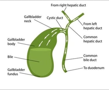
BILE JUICE :
· It is secreted by liver.
· It is passed into the duodenum by duct.
· It passes from the hepatic duct along with Cystic duct to the gallbladder ,where it is stored.
· Bile has a PH of 8
· Total secreted daily 500 -1000ml.
COMPOSITION OF BILE :
· Water
· Mucus
· Mineral salts
· Bile salts
· Bile pigments, mainly bilirubin
· Cholesterol
FUNCTION OF BILE JUICE :
(A)EMULSIFICATION OF FAT :
Bile contains bile salts that break large Fat Globules into tiny droplets increasing the surface area for enzyme like lipase to act.
(B)AIDS IN DIGESTION AND ABSORPTION:
Bile helps in the digestion and absorption of fat soluble vitamins (A,D,E,K).
(C)NEUTRALIZE STOMACH ACID :
Bile is A alkaline fluid and help neutralize the acidic chyme from the stomach creating an optimal environment for intestinal enzymes.
(D)EXCRETION OF WASTE :
Bile carries waste products like bilirubin and excess cholesterol out of Body via faeces.
(2)ABSORPTION :
It absorbs nutrients like glucose, amino acid, fatty acid, vitamins and minerals into the bloodstream through structures called Villi and Micro Villi.
(3)TRANSPORT :
It moves food along with its length by Peristalsis pushing it towards the large intestine.
(4)IMMUNE DEFENSE :
Contain lymphoid tissues that help defend against harmful microbes.
(5)HORMONE SECRETION :
It Release hormones like secretin and Cholecystokinin which regulate digestion by stimulating the pancreas and gall.
(6)WATER AND ELECTROLYTES BALANCE :
It absorbs a large amount of water and electrolytes helping maintain the body fluid balance.
(7)DETOXIFICATION :
It can help breakdown some toxins and prevent harmful substances from entering the bloodstream.
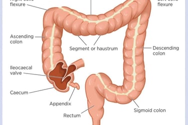
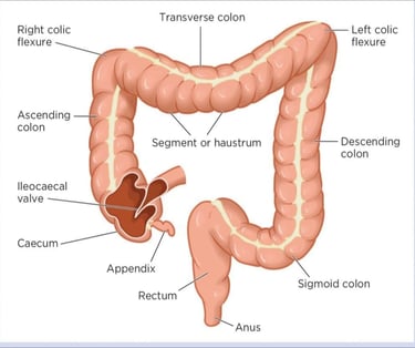
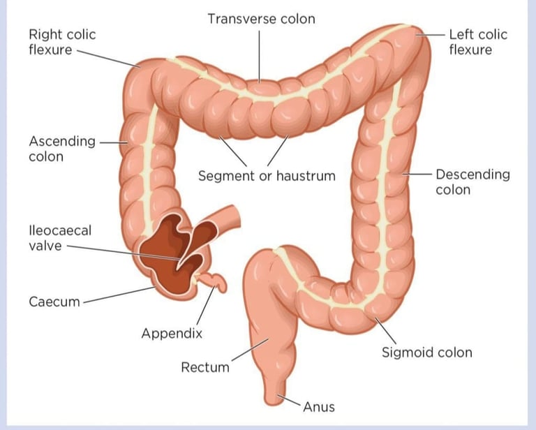
LARGE INTESTINE :
Large intestine is a wide muscular tube about 1.5 meter long.
It is divided into several parts :
1 CAECUM
2 APPENDIX
3 COLON
(A)ASCENDING COLON
(B)TRANSVERSE COLON
(C)DESCENDING COLON
(D)SIGMOID COLON
4 RECTUM
5 ANAL CANAL
(1)CAECUM :
· It is the first part of large intestine.
· It is about 5 to 8 cm long.
· It is a blind pouch that receive Chyme from the small intestine through ileocecal valve.
(2)APPENDIX :
A small finger like projection attached to the Caecum (has immune function but no digestive role).
(3)COLON :
(A)ASCENDING COLON :
Travels upward on the right side of the abdomen.
(B)TRANSVERSE COLON :
Crosses the abdomen from right to left.
(C)DESCENDING COLON :
Travels down the left side.
(D)SIGMOID COLON :
it is an S shaped segment that connects to the Rectum.
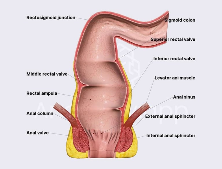
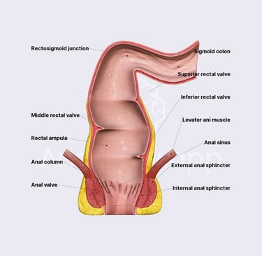
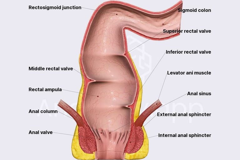
(4)RECTUM :
It is about 20 cm long.
It starts from Sigmoid Colon and ends in anal canal.
It store faeces before they are expelled.
(5)ANAL CANAL :
· It is about 2-3 cm long.
· It has two Sphincters.
· Internal Sphincter is under involuntary control .
· External Sphincter is under voluntary control.
· Anus is the opening through which faces are excreted ,controlled by internal and external anal sphincters.
STRUCTURE OF LARGE INTESTINE :
(A)MUCOSA :
· It consists of simple columnar epithelium and smooth muscles.
· Epithelium contains absorptive and goblet cells.
· The absorptive cells absorb water and goblet cells secrete mucus.
(B)SUBMUCOSA :
· It is made up of loose connective tissue that provides strength and flexibility.
· It contain blood vessels that supplies the submucosa with nutrients.
· It contain lymphatic vessels that helps in fluid balance and defence against infection.
· It also contain nerve plexus which controls secretion and blood flow to the intestinal wall.
It contain immune cells.
(C)MUSCLE LAYER :
It consists of external longitudinal muscles and an internal layer of circular muscles.
FUNCTION OF MUSCLE LAYER :
(1)PERISTALSIS :
Wave like movements that push faecal material forward.
(2)MASS MOVEMNT :
Powerful contractions that move waste towards the rectum.
(D)SEROSA :
· SEROSA of the large intestine is the outermost layer.
· It is made of a thin layer of connective tissue covered by a simple squamous epithelium.
· The serosa is the part of visceral peritoneum that lines the abdominal organs.
· Not entire large intestine is covered by serosa.
· In parts like ascending colon, descending colon, and parts of rectum outer layer is Adventitia, not serosa.
· In caecum ,transverse colon ,sigmoid colon, some parts of rectum a true serosa is present.
BLOOD SUPPLY :
Superior and inferior mesenteric artery supply the blood.
VENOUS DRAINAGE :
Superior and inferior mesenteric vein drain the blood.
NERVE SUPPLY :
Sympathetic supply is Via superior mesenteric plexus, inferior mesenteric plexus and hypogastric plexus.
Para sympathetic supply is from vagus nerve and splenic nerve.
FUNCTION OF LARGE INTESTINE :
(1)ABSORPTION :
Water and electrolytes (like sodium and chloride) are absorbed from the remaining indigestible food matter.
This process helps form solid faeces from liquid chyme.
(2)FORMATION AND STORAGE OF FAECES :
Large intestine compacts waste products into faecal material.
Faeces are stored in the rectum until defecation.
(3)BACTERIAL FERMENTATION :
Large intestine houses a large number of bacteria.
These bacteria ferment undigested carbohydrates to produce gases.
(4)VITAMIN PRODUCTION :
Gut bacteria synthesize some vitamins such as vitamin K And Vitamin B.
(5)IMMUNE FUNCTION :
Large intestine play role in immune defence containing lymphoid tissues that helps protect against harmful pathogens.
(6)DEFECATION :
· Mass movements push faecal material from the sigmoid colon into the rectum.
· This Results in distension of the rectal wall stimulates stretch receptors which initiate a defaecation reflex that empties the rectum.
· The pressure along with voluntary contractions of the diaphragm , abdominal muscles and para sympathetic stimulation open the internal sphincter and faeces are expelled through the anus.
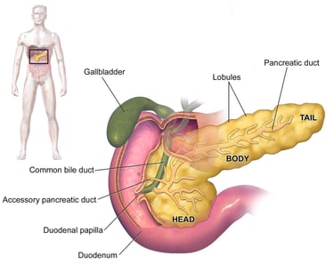
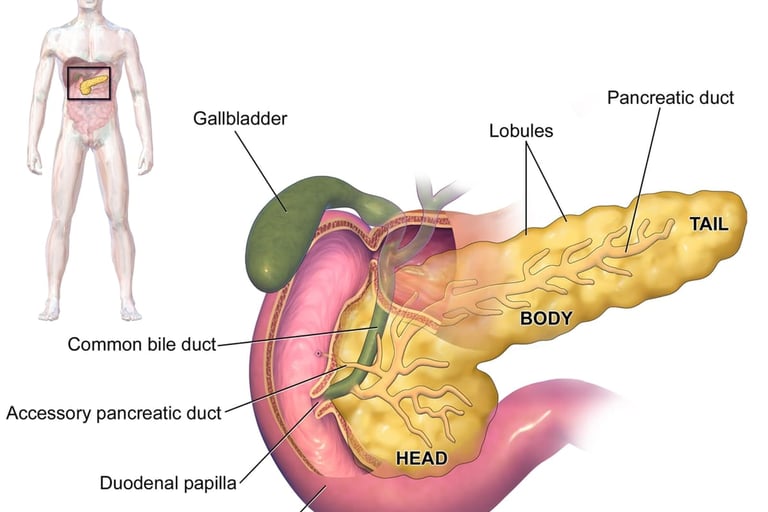
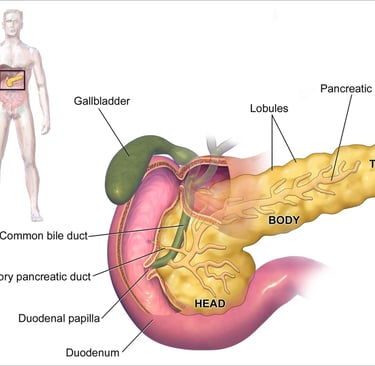
PANCREAS :
PANCREAS IS A GREYISH PINK COLOURED GLAND.
Its length is about 12 – 15cm
Weight -60gm
LOCATION : Lies behind the stomach across the posterior abdominal wall.
PARTS OF PANCREAS :
(A)HEAD :
Nested in the course of the duodenum.
(B)NECK :
Short part b/w head and body.
(C)BODY :
Extend across the midline.
(D)TAIL :
Touches the spleen.
Pancreas is both a endocrine and exocrine gland.
FUNCTION OF PANCREAS :
(1)EXOCRINE GLAND :
· Pancreas produce and secrets digestive enzymes and bicarbonate into the duodenum.
· These enzymes help break down of protein, carbohydrates and fats.
· Bicarbonates neutralize stomach acid.
· Pancreas consists of large no. of lobules.
· The secretory cells are present in the lobules.
· These pass their secretion into small ducts.
· This small duct passes into large duct.
· Larger duct is called pancreatic duct.
· Pancreatic ducts join the common bile duct from liver and gall bladder.
· It enters the duodenum as a common duct called hepatopancreatic duct.
(2)ENDOCRINE FUNCTION :
Pancreas contain cluster of pancreatic islets called (ISLET OF Langerhans).
It contain three types of cells.
(A)ALPHA CELLS :
· It secrete glucagon.
· Glucagon raises the blood glucose level in case of hypo-glycemia/exercise.
(B)BETA CELLS :
· It secrete insulin.
· Insulin regulates blood glucose level by promoting the uptake of glucose into cells.
· Insulin lowers blood glucose level by facilitating the entry of glucose into the cells, where it cab be used for energy.
(C)DELTA CELLS :
· It secrete somatostatin.
· Somatostatin hormone inhibits the release of insulin and glucagon.
· Hence, maintain glucagon and insulin secretion.
BLOOD SUPPLY :
Arterial supply to pancreas is supplied by splenic and mesenteric arteries.
Splenic and mesenteric veins drain the venous blood and joins portal vein.
NERVE SUPPLY :
Para sympathetic supply increase the secretion of pancreatic juice.
Sympathetic stimulation decrease the secretion of juices.
LIVER :
· Liver is a large, reddish brown organ.
· Liver is the largest gland in the body.
· Its weight is 1.4 kg.
LOCATION :
Upper right quadrant of the abdomen.
ASSOCIATED ORGANS :
SUPERIOR : Diaphragm and anterior abdominal wall.
INFERIOR : Stomach, bile duct ,duodenum , adrenal gland , kidney.
POSTERIOR : Oesophagus, inferior vena cava , aorta , gall bladder , vertebral column .
LATERAL : Lower ribs and diaphragm.
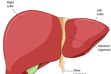
LOBES :
Liver has 2 main lobes.
· Right lobe (larger)
· Left lobe (smaller)
Right lobe and a smaller left lobe are separated by the falciform ligament.
Two additional lobe :
· Caudate lobe
· Quadrate lobe
STRUCTURE OF LIVER :
CAPSULE
The liver is covered by a thin ,strong connective tissue layer called Glissons capsule.
This capsule is also covered by the peritoneum.
INTERNAL STRUCTURE :
· The liver is made up of many functional units called lobules.
· Each lobule contain rows of hepatocytes.
· Liver has larger spaces lined by endothelium called sinusoids , through which blood passes.
· Sinusoids are lined by kuffer cells.
· Kuffer cells destroy worn out white and red blood cells ,bacteria and other foreign body in the blood.
· Hepatocytes produce bile.
· Bile flows through tiny canals to bile ducts and eventually to the gall bladder and small intestine.
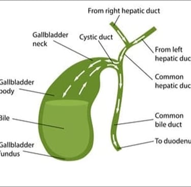
· Right and left hepatic duct unite and form common hepatic duct.
· Common hepatic duct join cystic duct from the gall bladder.
· Hepatic duct and cystic duct combine together to form common bile duct.
PORTAL FISSURE :
· Posterior surface of liver is known as portal fissure.
· Various structure enter and leave the liver.
· Hepatic artery enters carrying arterial blood.
· Portal veins enter and leave.
· Lymph vessels leave the liver, draining lymph to abdominal and thoracic nodes.
· Nerve fibres sympathetic and para sympathetic enter here.
BLOOD SUPPLY :
· Arterial supply is by hepatic artery.
· Portal veins bring nutrient rich blood from venous drainage is by hepatic vein into inferior hepatic vena cava.
FUNCTION OF LIVER :
(1)CARBOHYDRATE METABOLISM :
· Liver helps in maintaining the normal blood glucose level by storing glucose as glycogen(glycogenesis) and breaking its down when needed (glycogenolysis).
(2)FAT METABOLISM :
· Convert excess carbohydrates and protein into fat (lipogenesis).
· Produce cholesterol and lipoproteins.
· It convert fat into fatty acid.
(3)PROTEIN METABOLISM :
· The liver break protein into amino acids so that they can be used for ATP production.
· Convert, resulting toxic ammonia into urea for safe excretion through urine.
(4)DETOXIFICATION :
Breaks down and remove toxins, drugs, alcohol from the blood.
(5)BILE PRODUCTION AND SECRETION :
· Produce Bile which help digest fat in small intestine.
· It also helps in absorption of fat in small intestine.
(6)STORAGE :
Act as a storage for substances secreted :
· Glycogen
· Vitamins (A,D,E,K AND B12)
· Minerals (IRON,COPPER)
(7)HELPS IN IMMUNITY :
· Kuffer cells are specialised immune cells that destroy bacteria and wornout cells.
· Helps in bodies defence against infection.
(8)SYNTHESIS OF BLOOD CLOTTING FACTORS :
Produce fibrinogen and prothrombin proteins, necessory for blood clotting.
(9)HORMONE REGULATION :
Metabolise and regulate level of hormones like insulin, thyroid hormone and sex hormone.
(10)PRODUCTION OF HEAT :
Liver uses some amount of energy and helps in production of Heat in body.
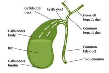
GALL BLADDER :
Gall bladder is a small pear shaped organ located just under the liver on the right side of the abdomen.
STRUCTURE OF GALL BLADDER :
(1)FUNDUS :
The broad rounded End.
(B)BODY :
The main central part.
(C)NECK :
Narrow part that connects to the cystic duct.
DUCT SYSTEM :
· Cystic duct from gallbladder join common hepatic duct from the liver combines to form common bile duct.
· The common bile duct carries bile to the small intestine.
Gall bladder has three layers :
(1)MUCOUS MEMBRANE :
· It is the inner mucous membrane.
· It is composed of Columnar epithelium cells, which secrete mucin and rapidly absorb water and electrolytes.
(2)MUSCULAR LAYER :
· This consists of Smooth muscle fibres.
· Contracts to squeeze bile out of the gall bladder when needed.
(3)SEROSA :
· It is the outermost layer that covers the portion that are not directly attached to liver.
· it is a thin layer of mesothelium and connective tissue.
· This layer is distinct from the Adventitia.
Blood Supply :
· Hepatic duct supply and cystic duct supply arterial blood.
· Cystic vein drain venous blood further join portal vein.
NERVE SUPPLY :
Sympathetic and Parasympathetic nerve fibres convey nerve impulses.
FUNCTION OF GALL BLADDER :
(1)STORAGE OF BILE :
Liver produce bile, gall bladder stores bile instead of letting it go constantly drip into the small intestine.
(2)CONCENTRATION OF BILE :
Gall bladder removes water and electrolytes from the bile.
This make Bile thicker and more concentrated, making it more effective for digestion.
(3)RELEASE OF BILE :
· When we eat oily food a hormone called cholecystokinin is released.
· Cholecystokinin signal the gallbladder to contract and release bile into the small intestine through the common bile.
(4)AID IN FAT DIGESTION AND ABSORPTION :
· Bile helps emulsify fat- breaking large fat droplets into tiny ones.
· This help lipase to work, making fat digestion faster and more efficient.
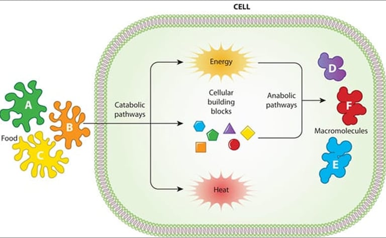
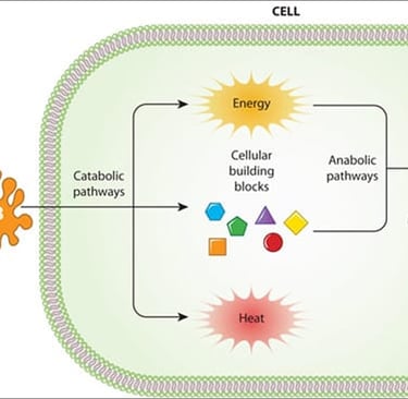
METABOLISM :
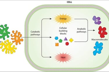
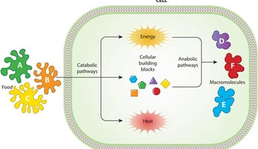
· Metabolism refers to All chemical reactions that happen inside the body.
· These reactions involve breaking down nutrients to produce energy.
· Building Important molecules like protein and DNA molecules.
TYPE OF METABOLISM :
(1)CATABOLISM (BREAKING DOWN):
· Large molecules are broken on into smaller ones, it release energy example breaking glucose during digestion to release energy in the form of ATP.
(2)ANABOLISM :
· Small molecules are built up to layer complex ones.
· Consumes energy .
· Example building muscle protein from amino acid.
CARBOHYDRATE METABOLISM :
· Breaks down sugar and starch into glucose.
· Glucose is used to produce ATP.
· Extra glucose is stored as glycogen.
FAT METABOLISM :
· Fats are broken into fatty acids And Glycerol used for energy in future.
· Used during fasting and exercise.
PROTEIN METABOLISM:
· Proteins are broken down into amino acid.
· Used to build new proteins, If needed converted to glucose for energy.
MAJOR METABOLISM OGANS :
· Liver
· Muscles
· Brain
· Adipose tissue
ENERGY PRODUCTION :
· All metabolism Aims to create ATP.
· ATP provides energy for body activities such as :
Nerve impulse conduction
Muscle contraction
Cell division.
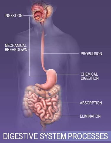
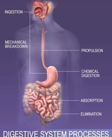
DIGESTIVE SYSTEM PROCESS:
(1)INGESTION :
· It is the first step.
· This is a process of taking food inside the mouth for its journey through the digestive system.
· Food enters the mouth where chemical and mechanical digestion of food begin.
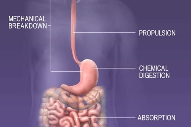
(2)PROPULSION :
· It is the second step .
· In the step food moves forward into the Oesophagus through peristalsis.
(3)DIGESTION :
· The breakdown of food particles into smaller molecules so that they enter the cell is called digestion.
Digestion takes place by two ways:
(A)MECHANICAL DIGESTION :
It is done by person himself by Mastication (chewing).
(B)CHEMICAL DIGESTION :
In this food is mixed with saliva, its enzymes and other digestive juices which break food into smaller molecules.
(4)ABSORPTION :
· Nutrients are Absorbed into the blood stream through intestinal walls.
· Water and electrolytes are absorbed in the large intestine of digestive tract.
(5)ELIMINATION :
Waste is expelled from the body through rectum and anus during defecation.