UNIT -7
NERVOUS SYSTEM
Nervous system is made up of a vast number of cells called neurons.
There are about 100 billions of neurons in the brain.
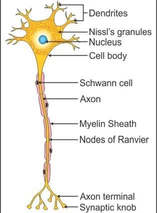
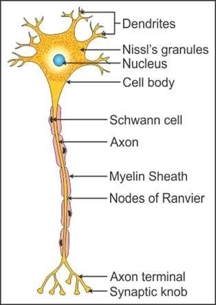
Structure of a Neuron:
Each Neuron consists of a cell body and its process, one Axon, and many Dendrites.
These are thread like structures called nerve fibers.
The cell body contains nucleus ,cytoplasm and various organelles found in cells like mitochondria and Golgi apparatus.
Neuron cell cannot divide.
They need continuous Supply of oxygen for their survival.
They can synthesize energy only from glucose.
CELL BODY :
The nerve cells vary in shape and size.
They are too small to be seen by the naked eyes
Cell bodies form gray matter.
These are present at periphery in the brain and Centre of the spinal cord .
Cell bodies collectively form a group called Nuclei in the central nervous system and it is called Ganglia in the peripheral nervous system.
AXON AND DENDRITES :
Axon and dendrites are the branches which extend from a tapered portion of cell body .
One Neuron has one Axon but many Branches of Dendrites .
Nerve fiber is a term used for neuron process ( Axon and Dendrites )
Most nerves contain bundle of both sensory and motor nerves .
Axon and Dendrites form White matter in the Nervous system.
AXON :
Axon of a Neuron is a single process that extends from a tapered portion of cell body .
One neuron has only one axon.
Axons conduct impulses away from the cell body.
An axon is a long thin cylindrical projection.
These are 100 centimeter long.
Axons in group are called Tracts.
Axons are found deep in the brain and at periphery of the spinal cord.
STRUCTURE OF AN AXON :
The axon contains the endoplasmic reticulum, mitochondria and microtubules .
Its cytoplasm is called Axoplasm.
It is surrounded by a plasma membrane known as Axolemma.
The Axon is surrounded by a Myelin Sheath.
Schwann cells are present in the inside of an axon body.
They are wrapped around the axon .
Microscopic gaps in the Sheath, between adjacent Schwann cells are called nodes of Ranvier .
Myelin Sheaths and gaps are important in the proper conduction of impulses along nerve fibers.
Outer layer of Schwann cells is called Neurilemma.
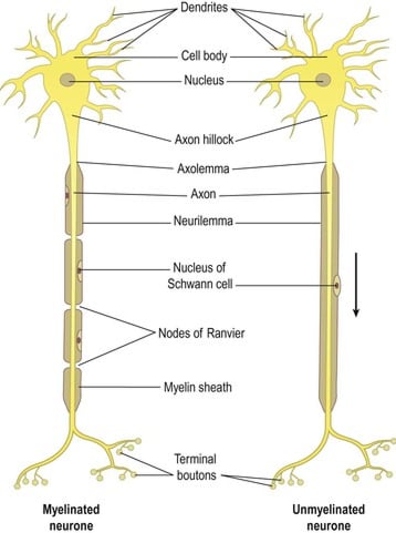
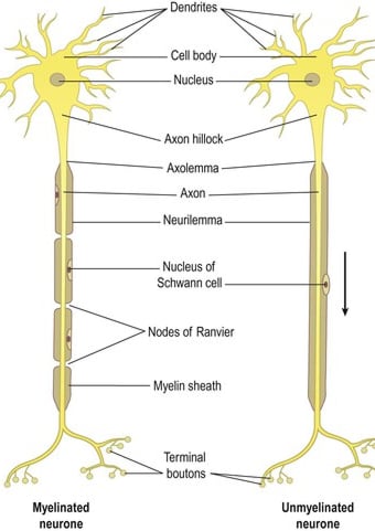
Axon of neurons are surrounded by a multi lipid layer and a protein covering called Myelin Sheath.
Axon with this covering are myelinated and those without it are unmyelinated.
Spread of nerve impulses transmission is lower in Non Myelinated fibers.
DENDRITES :
Dendrites’ are the branches arise from cell body.
Dendrites conduct impulses to the cell body of a neuron.
They are usually short tapering and highly branched.
Dendrites are not Myelinated.
ACTION POTENTIAL :
A Nerve impulse is a wave of electrical fluctuation that travels along the plasma membrane.
Neuron maintain a difference in the concentration of ions across their membrane.
There is slightly excess of positive ions on the outside of the membrane .
There is slightly excess of negative ions inside.
This result in a difference in electrical charge across the plasma membrane called membrane potential.
A membrane that exhibit membrane potential is said to be polarized.
Membrane potential maintained by a Non conducting neuron plasma membrane is called Resting Membrane Potential.
Principal ions involved are :
(1)Sodium (Na+) the main extracellular cation.
(2)Potassium (K+) the main intracellular cation.
Excitation of neuron occurs when a stimulus triggers opening of additional sodium channel which permit more sodium ion to enter into the cell.
As excess of positive ions outside the plasma membrane decrease membrane potential is reduced it is known as depolarization .
It creates an action potential .
Depolarization is a very rapid process that enables conduction of nerve impulses along the entire length of neuron in few milliseconds.
Once the peak of the action potential is reached the membrane potential begins to move back towards the resting potential the process is called repolarization .
Refractory Period is brief period in which membrane resists Restimulation.
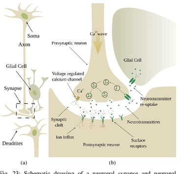
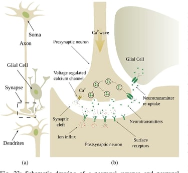
SYNAPSIS :
The connection between the two neurons is called synapse.
One neuron is connected to another neuron not Anatomically but functionally.
One neuron pass information to another neuron with the help of neurotransmitter and synaptic vesicles.
STRUCTURE OF SYNAPSE :
(1) PRESYNAPTIC CLEFT :
It contain Neurotransmitters, Mitochondria and other cell organelles.
(2) POST SYNAPTIC CLEFT :
It is a site for receiving neurotransmitters.
(3) SYNAPTIC CLEFT :
A space between pre synaptic and post synaptic Cleft.
FUNCTION OF SYNAPSIS :
The main function of synapse is to transmit the nerve impulses.
It occurs through two parts .
(1) Electrical Transmission :
The membrane of a Two cell actually touch and share a potential.
This allows the action potential to pass directly from one membrane to another .
(2)Chemical Transmission:
From the pre- synaptic vesicles of neuron a chemical substance release called Neurotransmitter that binds to the plasma membrane of Post synaptic neuron .
(3)Transmission of Neurotransmitters :
One neuron influence the activity of other neuron by releasing a neurotransmitter from the synaptic vesicles
There are two types of Neurotransmitters .
(1)Excitatory Neurotransmitters.
(2)Inhibitory Neurotransmitters.
(1)Excitatory Neurotransmitters :
Examples are:
Acetylcholine
Dopamine
Norepinephrine
(2)Inhibitory Neurotransmitters :
· Examples are :
Serotonin
GABA
Glycine
NERVES :
Nerves are the Messenger that carries the message to and from the brain.
TYPE OF NERVE :
(1)Sensory Nerve OR Afferent Nerve :
These transmit sensory impulses from receptors in the skin, sense organs, and muscles into the central nervous system.
Common/ Somatic senses convey impulses for the sense of pain, temperature ,touch vibration .
Special senses convey impulses for sense of Taste and Smell, Sense of vision, Sense of Hearing and sense of Touch.
(2)MOTOR OR EFFERENT NERVE :
These convey impulses from the central nervous system to the Effector organs.
These include two type of nerve:
(1) SOMATIC NERVE :
Involved in contraction of Skeleton Muscles.
(2)AUTONOMIC NERVES :
Involved in contraction of smooth muscles, cardiac muscles via cranial and spinal nerves.
(3)MIXED NERVES :
Some are sensory and some are motor nerves in the spinal cord arrangement.
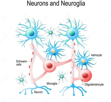
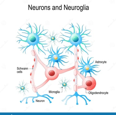
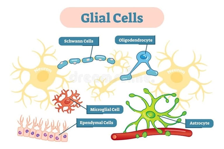
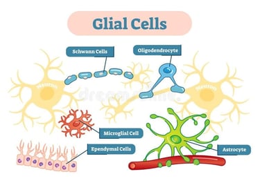
NEUROLGIA :
These are smaller than neurons, these are Non Excitable glial cells.
These do not conduct electrical impulses.
These are the supporting cells provide nourishment and oxygen to the neurons .
These are of four types:
(1) Astrocytes:
These are star shaped cells with many process.
Function of Astrocytes :
They participate in the metabolism of neurotransmitters.
They maintain proper balance of potassium ions for generation of nerve impulses.
They participate in brain development.
They help to form blood brain barrier which regulates entry of substances into the brain .
They provide a link between your Neurons and Blood Vessels.
(2)Oligodendrocytes:
These are the most common glial cells in central nervous system .
They form a supporting network by twinning around neurons.
Main function:
They provide a lipid and a protein wrapping called a myelin Sheath.
(3)Microglia :
These are phagocytic neuroglia derived from monocytes .
MAIN FUNCTION :
They protect the central Nervous system from disease engulfing invading microbes and clearing away debris.
(4) Ependymal cells:
These are Epithelial cells.
They Range in shape from cuboidal to columnar.
Function:
They form cerebrospinal fluid and assist in its circulation.
CENTRAL NERVOUS SYSTEM :
Central Nervous system consists of brain and spinal cord.
Structure of Brain and Spinal Cord :
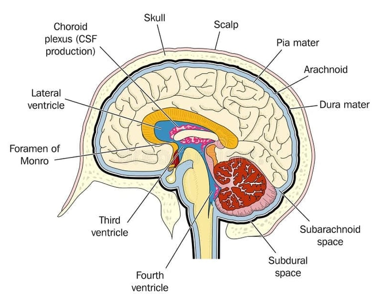
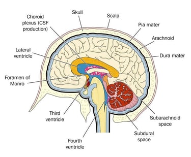
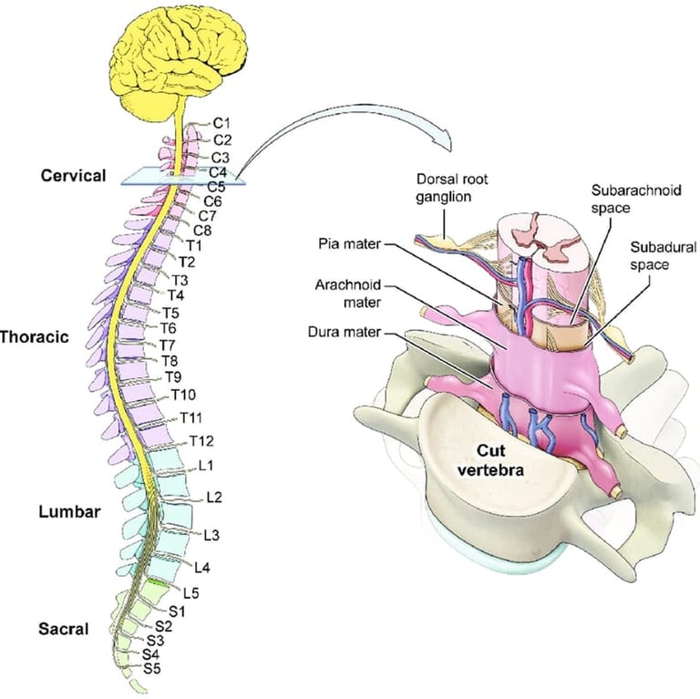
Brain and Spinal cord are surrounded by Meninges which protect the delicate nerve structure.
Meninges :
(1) Dura-mater
(2) Arachnoid mater
(3) Pia-mater
(1)Dura-mater :
It is a dense and Tough layer .
It consists of two layers.
one outer layer lines the skull.
The Inner layer is united with it ,Except where the Venous sinuses are formed .
The Falx cerebri lies between the two cerebral hemisphere, its upper border forms the superior longitudinal sinus receiving venous blood from the brain.
Its lower margin form inferior longitudinal ,drains the falx cerebri.
Tentorium Cerebelli separates the cerebrum from the cerebellum.
(2)Arachnoid mater :
Arachnoid mater is the delicate serous membrane.
It lies between the Dura-mater and Pia-mater.
It consists of delicate collagen fibers and some elastic fibers.
Between the dura-mater and Arachnoid mater there is a thin subdural space which contain interstitial fluid called cerebrospinal fluid.
(3)Pia-mater :
Pia-mater is a thin vascular membrane.
It adheres to the surface of the spinal cord and brain.
It contains bundle of collagen fibers and some fine elastic fiber’s.
It contain many blood vessels that supply nutrients and oxygen to the spinal cord.
VENTRICLES OF BRAIN :
The fluid circulate within the Brain and Spinal cord through the cavities .
These cavities are:
Right and left lateral ventricles
Third Ventricle
Fourth Ventricle
(1)Lateral Ventricles :
These cavities are located in a hemisphere of the Cerebrum.
These are present on each side of the median plane just below the Corpus Callosum .
The Septum Lucidum separates them.
The Interventricular Foramina helps to communicate with Third Ventricle.
(2)The Third Ventricle :
This is in narrow cavity at midline superior to the hypothalamus.
It is present between the right and left halves of the thalamus, just below the lateral ventricles.
The cerebral Aqueduct of Mid Brain helps to communicate with the Fourth Ventricle.
(3)Fourth Ventricle :
It is a diamond shape.
It lies between a brain stem and the Cerebellum.
The Foramina is present in its roof .
Help in communication with the Sub-Arachnoid space.
Cerebrospinal Fluid (CSF) :
Cerebrospinal fluid formation occurs by separation of fluid from the blood in Choroid plexus.
Choroid Plexus are network of capillaries that enter into the lateral ventricles, from each lateral ventricle fluid seeps through an opening the interventricular foramen into the 3rd ventricle.
Then through Aqueduct of Sylvius into the 4th ventricle.
The fluid circulates in the Sub-Arachnoid space then is Absorbed into the venous blood through the arachnoid villi .
CSF IS absorbed as rapidly as it is formed by the choroid plexus at the rate of about 20 ml per hour ( total 480 /day ) because the rate of formation and reabsorption are same in the pressure of CSF.
Formation, circulation and reabsorption of cerebrospinal fluid is normally constant .
CSF Pressure may be measured using a Vertical tube Attached to the Lumbar puncture needle.
It remains constant at about 10 cm h2o when the individual is lying on his side and about 30 cm H2O when sitting up.
Properties of CSF :
CSF is A alkaline fluid.
Its specific gravity is 1.005
CSF is a clear fluid.
It contains :
Water
Minerals
Glucose
Plasma Proteins, small amount of albumin and globulin
Creatinine
Urea
A few leukocytes.
FUNCTION OF CEREBROSPINAL FLUID :
The CSF maintains the Homeostasis .
(1)MECHANICAL PROTECTION :
The fluid serve as a shock observing medium it protects the delicate tissue of the brain and spinal cord.
(2)CHEMICAL PROTECTION :
CSF provides an optimal chemical environment for neural signals. Even the slight change in CSF Production disrupt action potential.
(3)CIRCULATION :
CSF is a medium for exchange of nutrients and waste products between blood and neurons tissue.
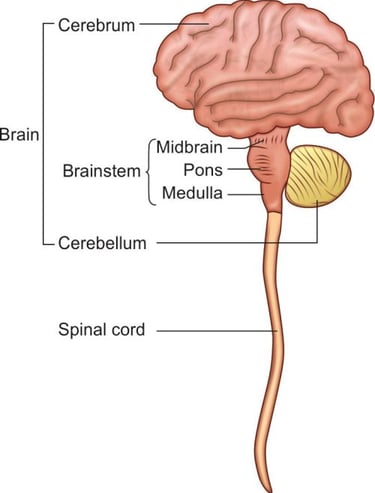
BRAIN :
It lies within the cranial cavity .
The parts of brain are :
CEREBRUM
MIDBRAIN
PONS
MEDULLA OBLONGATA
CEREBELLUM
(1)CEREBRUM :
The cerebral is made up of two Cerebral Hemisphere the right and left hemisphere.
These are incompletely separated from each other by the median longitudinal Fissure.
Two Hemisphere are connected to each other by the Corpus Callosum.
Superficial layer of the cerebrum is grey matter and is called cerebral cortex.
Deep to the cortex lies the cerebral white mater.
Cerebral cortex enlarges and shows Deep furrows of varying depth.
Folds are called gyrus or convulsions.
Deepest grooves between the folds is known as fissures.
Shallower grooves between folds are termed as Sulci.
The most prominent fissure is longitudinal fissure.
It separates the right and left hemisphere.
Each cerebrum of the hemisphere is divided into lobes .
The lobes are :
(1) Frontal LOBE
(2) PARIETAL LOBE
(3)TEMPORAL LOBE
(4)OCCIPITAL LOBE
NAME AND LOCATION OF PROMINENT CEREBRAL FISSURES ARE FOLLOWING :
(1)LONGITUDINAL FISSURE :
Deepest groove in the cerebrum divides the cerebrum into two hemispheres.
(2)CENTRAL SULCUS :
A groove between the frontal and Parietal lobe.
(3)LATERAL FISSURE :
A deep groove between the temporal lobe below, the frontal and Parietal Lobe Above.
(4)PARIETO-OCCIPITAL FISSURE :
Groove that separates the occipital lobe from the two Parietal lobes.
FUNCTION OF CEREBRUM :
The cerebrum is the seat of intelligence, memory, reasoning ,thinking speaking, reading, writing.
Sensory Perception includes the Perception of pain, Temperature, Touch, Sight, hearing, taste and smell.
It stimulates and control the skeleton muscle contraction.
FUNCTIONAL AREAS OF CEREBRUM :
MOTOR AREA OF CEREBRUM :
(1) PRIMARY MOTOR AREA :
Located in the pre central gyrus of the frontal lobe.
This area controls voluntary contraction of muscles.
(2)MOTOR SPEECH AREA :
Production of speech occurs in Broca’s area, it is located in left frontal lobe superior to central Sulcus, controls muscle movement necessary for speech.
SENSORY AREA OF THE CEREBRUM :
(1)GENERAL SENSORY AREA :
It is present in the post central gyrus of each parietal lobe .
This area is for touch, pain and temperature.
(2)PRIMARY VISUAL AREA :
It is located on the medial surface of the occipital lobe .
The optic nerve pass from the eye to this area, which receive and interpret the impulse as visual impression.
(3)PRIMARY AUDITORY AREA :
Located in the superior part of the temporal lobe.
It interpret the basic Characteristics of sound such as pitch and rhythm.
(4)PRIMARY GUSTATORY AREA :
Located in the base of the post central gyrus present in the Parietal cortex .
It receives impulses related to Taste.
OTHER AREAS OF CEREBRUM :
It is embedded in the mass of white matter of each cerebral hemisphere.
There are certain areas of grey matter.
It influence tone and posture, integrate and coordinate the main voluntary muscle movement.
(2)THALAMUS :
It is concerned with the receptor of sensory impulses.
It regulates the action for sensation and movement.
(3)HYPOTHALAMUS :
Function performed by Hypothalamus are:
Body temperature maintenance.
Hunger and thirst.
Emotional reactions example pleasure, fear, rage.
Autonomic nervous system maintenance.
Appetite.
Sexual behaviour.
Biological clock, example sleeping and waking cycle.
BRAIN STEM
MIDBRAIN
PONS
MEDULLA OBLONGATA
(1)MIDBRAIN :
Midbrain forms the upper part of the brain stem.
It runs the Cerebral Aqueduct connecting the Third and Fourth ventricle.
Midbrain contains centre for the control of balance and movements of the eyes.
(2)PONS :
The Pons form the middle portion of the brain stem.
It contains some ascending and descending pathways as the midbrain.
It connects the brain to the spinal cord and medulla oblongata.
FUNCTION OF PONS :
(1)SENSES :
The Pons help in hearing and balance.
(2)MOVEMENT :
The Pons help with eye and facial movement and the ability to move parts of the mouth.
(3)BREATHING :
The Pons help in control of rhythmic breathing.
(4)SLEEP :
The pons help generate sleep.
(3)MEDULLA OBLONGATA :
It is 2.5 centimetre long .
It links the pons with the spinal cord .
Medulla Oblongata contain certain vital centre which are :
(1)Respiratory centre controls the respiratory rate.
(2) Cardiovascular centre.
(3) Vasomotor centre.
(4) Reflex centre of vomiting, coughing, sneezing and swallowing.
· Damage to the Medulla oblongata data can be life threatening
· Symptoms include breathing problems, loss of muscle control and balance problems.
CEREBELLUM :
Cerebellum is the second largest portion of the brain.
It is posterior to the medulla oblongata and pons, inferior to the cerebrum.
FUNCTION :
It maintain the posture of the body .
Maintenance of balance.
It play role in muscular coordination.
Cerebellum helps in skilled motor activities from catching a base ball to dancing.
Damage to the cerebellum causes disturbance of posture and muscle tone, staggering gate etc.
Movement is very incoordinate.
All voluntary movement is low.
SPINAL CORD :
The spinal cord begins at the medulla oblongata.
It emerge from the Foramen Magnum and end between first and second lumber vertebra.
The cord is about 45CM long.
It is divided in front by a deep anterior fissure and A narrow one at the back.
Spinal cord is linked with brain as well as with our body parts.
The nerve impulses that arise from the brain and reach the various organs and tissues descend through the spinal cord sensory nerves.
Some activities of the spinal cord are independent of the brain that is spinal reflexes.
Spinal cord contains grey matter inside and white matter outside.
SPINAL REFLEXES :
Reflexes are fast predictable automatic responses to change in the environment.
The simplest kind of pathway is known as reflex arc.
Reflex arc includes the following functional component.
(1)SENSORY NEURON :
Nerve impulses propagate from the receptor to the axon terminal of the sensory neuron which is located in grey matter of the spinal cord or brain stem.
(2)MOTOR NEURON :
The part of the body that responds to the motor nerve impulses such as muscle/gland is the effector.
Its action is called reflex.
If the effector is smooth muscle, cardiac muscle or gland the reflex is an autonomic reflex.
PHERIPHERAL NERVOUS SYSTEM :
It consists of :
31 pairs of spinal nerves.
12 pairs of cranial nerves .
The Autonomic nervous system.
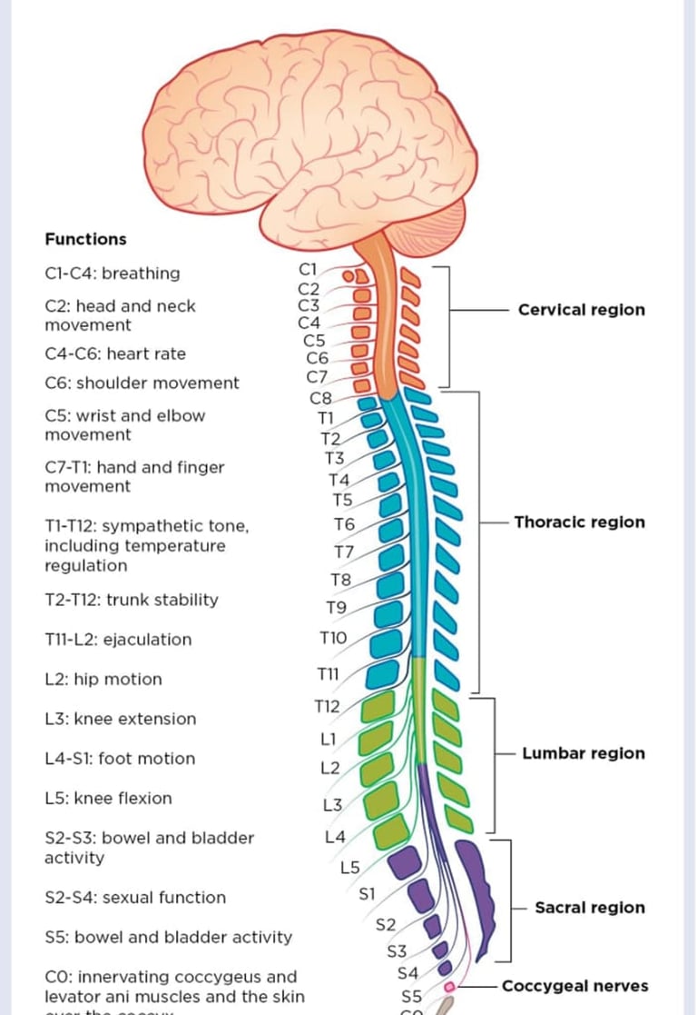
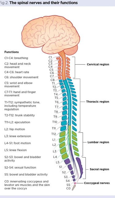
(1)SPINAL NERVES :
31 pairs of spinal nerves are named and numbered according to the region and level of the vertebral column.
8 pairs of cervical nerves C1 to C8
12 pairs of thoracic Nerves T1 to T12.
5 pairs of Lumbar nerves L1 to L5.
5 pairs of sacral nerves S1 to S5 .
1 pair of coccygeal nerves.


(2)CRANIAL NERVES :
There are 12 pairs of cranial nerves.
(1)OLFACTORY NERVE (SENSORY) :
• The olfactory nerve carry information about sense of smell .
• It passes through olfactory foramina in the cribiform plate of ethmoid bone and ends in olfactory bulb .
• The olfactory tract extends in the temporal bone.
(2)OPTIC NERVE (SENSORY) :
• These are the nerves of sense of sight.
• It arises in the Retina of the eye passes through optic Chiasma, passes through optic Tracts and terminate in lateral nuclei of thalamus.
(3)OCULOMOTOR NERVE (MOTOR) :
• Fibres of the Oculomotor nerve originate in the midbrain.
• Autonomic fibres are also present, they extend to the intrinsic muscles of the eye which regulate the amount of light entering into the eye and aid in focusing on nearby objects.
(4)TROCHLEAR NERVE (MOTOR) :
• The Trochlear nerve originates in the midbrain.
• They extend to the superior oblique muscle of the eye.
• Trochlear nerve controls eye movement.
(5)TRIGEMINAL NERVE (MIXED) :
These are called trigeminal because they split into 3 branches. The 3 branches are :
• Ophthalmic nerve
• Auxiliary nerve
• Mandibular nerve It provides sensory and motor information of face and head. It helps in mastication.
(6)ABDUCENT NERVE (MOTOR) :
• Sixth pair of cranial nerves, supply the muscles concerned with the lateral movement of the eyeballs.
(7)FACIAL NERVE (MIXED) :
• The Facial nerve arise from part of Pons.
• Nerve arises from taste buds on anterior two third of the tongue to the taste perception area in cerebral cortex.
(8)VESTIBULOCOCHLEAR NERVE (SENSORY) :
(A)COCHLEAR BRANCH :
• Cochlear branch convey impulses associated with hearing.
• Cochlear nerve consists of dendrites in the organ of Corti in the cochlea in the inner ear.
(B)VESTIBULAR BRANCH :
• Convey impulses associated with equilibrium.
• It arises in semicircular canal and ends in pons and cerebellum.
(9)GLOSSOPHARYNGEAL NERVE (MIXED) :
• The sensory portion of nerve arises from taste birds on posterior 1/3 of the tongue and ends in medulla.
• Its main function is taste, Regulation of blood pressure.
• Its motor part originates in medulla.
• Its function is To secrete Saliva.
(10)VAGUS NERVE :
• Vagus nerve contain both sensory and motor fibres.
• Its sensory fibres supply the Pharynx, larynx, trachea, heart, lung, bronchi and oesophagus, stomach, small intestine and gall bladder.
• Motor fibres control the muscles involved in swallowing.
(11)ACCESSORY NERVE (MOTOR) :
• It is a motor nerve its fibres originate in the medulla.
• It controls swallowing movements.
(12)HYPOGLOSSAL NERVE (MOTOR) :
• Hypoglossal nerve originate in medulla.
• It supply muscle of tongue.
• It help in movement of tongue during speech and swallowing.
AUTONOMIC NERVOUS SYSTEM :
It is divided into two parts.
(1)SYMPATHETIC SYSTEM :
STRUCTURE :
• Sympathetic system Is a system which lie in front of vertebral column and connected with spinal cord by nerves fibres.
• It consists of a double chain of gangliovated cord extending from the base of the skull, lying in front of vertebral column to end in the pelvis opposite to the coccyx as the ganglion pair. The ganglia are arranged in following regions
• In the neck -3 pairs of cervical ganglia.
• In the chest -11 pairs of thoracic ganglia.
• In the spine- 4 pairs of Lumber Ganglia.
• In the pelvis- 4 pairs of sacral ganglia.
• In the coccyx - One Ganglion pair.
FUNCTION OF SYMPATHETIC NERVOUS SYSTEM :
• Sympathetic nerve supply to the muscles of heart, the involuntary muscles of all blood vessels and Viscera such as stomach and pancreas.
• it maintains the tone of all muscles including tone of voluntary muscles.
(2)PARA SYMPATHETIC SYSTEM :
• The 3rd 7 th 9 th and 10th cranial nerve are the autonomic nerve.
• These form when parasympathetic fibres pass out from the brain to the organs.
• By the means of 3rd cranial nerve oculomotor nerve fibers reach the circular muscle fibres of the Iris, stimulating the movements which determine the size of the pupil of the eye.
• By the Seventh facial Nerve and 9th glossopharyngeal nerve, Motor fibres reach at the Salivary gland.
• Vagus nerve is the largest autonomic nerve.
• It has wide distribution in body and send fibres to a number of Glands and organs.