UNIT 5
LYMPHATIC SYSTEM
UNIT - 5
LYMPHATIC SYSTEM
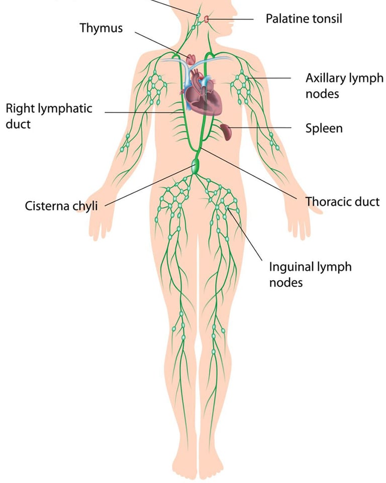
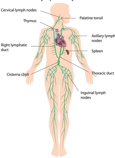
LYMPHATIC SYSTEM CONSISTS OF:
LYMPH
LYMPH NODES
LYMPH VESSELS
LYMPH OGRANS EG. SPLEEN, THYMUS GLAND
LYMPHOID TISSUE
BONE MARROW
LYMPH:
Lymph is a clear and watery appearing fluid inside the Lymphatic Vessels.
Lymph resembles Blood Plasma in composition.
Lymph is isotonic in nature. Total amount of Lymph - 2500 to 2800ml.
Lymph is a milky appearance fluid due to presence of fatty molecules inside it which gives it a milky appearance.
Lymph is a fluid that drains back into the Blood Circulation.
LYMPH VESSELS:
The Thickness of Lymph vessels is same as that of Veins. LAYERS OF LYMPH VESSELS:
Outer layer of fibrous tissue .
Middle layer of Smooth muscles.
Inner layer of Endothelium.
Lymph vessels contain Valves which allows one way flow of lymph.
The fluid moves forward by the contractions of muscles.
The Small lymph vessels join to form the larger vessels then join with other lymphatics to form larger vessels which merge to form main Lymphatic Trunks, The Right Lymphatic Duct and Thoracic duct.
From the Thoracic Duct the Lymph drains into Left Subclavian Vein.
ARTERIES
BLOOD CAPILLARIES
INTERSTITIAL SPACE
LYMPHATIC CAPILLARIES
LYMPHATIC VESSELS
LYMPHATIC DUCTS
SUBCLAVIAN VEIN (BLOOD PLASMA)
LYMPHATIC ORGAN :
(1) LYMPH NODE:
Lymph Node are small bean like bodies found in the lymphatic vessels.
They act as filters and are the sites where lymphocytes are formed.
Lymph Node lie in the Neck,Axilla, Thorax, Abdomen and Groin.
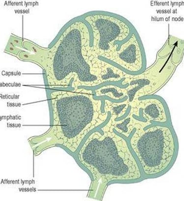
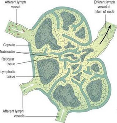
STRUCTURE OF LYMPH NODE :
Lymph Node is covered by a capsule of dense connective tissue.
The Capsular Extensions of Lymph Node are called Trabeculae.
The Trabeculae divide the Lymph Node into compartments, provide support and convey Blood Vessels into the Interior of a Lymph Node .
The Parenchyma of a Lymph Node is divided into 2 Regions.
Cortex And Medulla
Lymph flows through Lymph Node in one Direction.
It Enters through Afferent Lymphatic Vessels.
It Exit through Efferent Lymphatic Vessels.
Efferent Lymphatic Vessels emerge from one side of the Lymph Node at a slight depression called Hilus. Blood Vessels also Enter and Leave the Lymph Node at Hilus.
FUNCTION OF LYMPH NODE :
(1) FILTERATION :
Lymph Node filter lymph by having it, enter at one End and Exit at Another.
The Lymph Node filter foreign substances from lymph as it passes back towards the blood stream. Macrophages destroy some foreign substances by Phagocytosis.
(2) PROLIFERATION OF LYMPHOCYTES:
Lymph Node helps in maturation of T Lymphocytes.
And Transport them to other body parts after proliferation and maturation.
(3) HEMАTOPOIESIS:
THE Lymph Node Serves as Final Site of Maturation for some Lymphocytes and Monocytes that have migrate from Bone Marrow.
Hence, helps in formation of Blood Cells.
SPLEEN :
Spleen is the mass of largest single lymphatic tissue in the body.
Length - 12 cm
Width - 7 cm
Thickness - 22.5 cm
Location - Left Hypochondriac Region
Weight - 200 Mg
ORGANS ASSOCIATED WITH SPLEEN:
POSTERIORLY: DIAPHRAGM
ANTERIORLY: FUNDUS OF STOMACH
MEDIALLY : PANCREAS AND LEFT KIDNEY
LATERALLY: SEPARATED FROM 9TH, 10TH, AND 11TH RIBS AND THE INTERCOASTAL MUSCLES BY DIAPHRAGM.
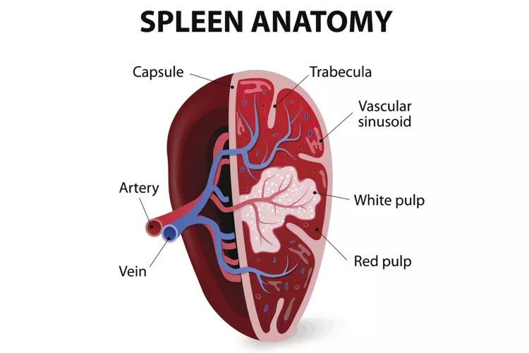
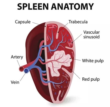
STRUCTURE OF SPLEEN:
Spleen is formed by Lymphoid tissue, Connective tissue and Several Blood cells.
It is Covered by a capable and few smooth muscle fibres.
The Parenchyma of spleen is formed by two different kind of Tissue called white pulp and red pulp.
The White pulp function in immunity as a site of B Cell proliferation into Antibody producing Plasma Cells. Red pulp causes Phagocytosis of bacteria and worn out damaged Red Blood Cells and Platelets.
FUNCTION OF SPLEEN :
(1) PHAGOCYTOSIS :
THE Spleen helps to destroy bacteria and worn out damaged Red Blood Cells.
(2) STORAGE OF BLOOD :
THE Spleen contain upto 350 ml of blood so that in response to Sympathetic stimulation can rapidly return a large volume of this blood to Circulation.
(3) IMMUNE RESPONSE:
THE B And T Lymphocytes are activated by Antigens produce antibodies and fight against infection.
(4) ERYTHROPOIESIS:
Spleen and Liver are site of fetal Blood Cells production and Spleen can also fulfill this function in adult at great time of need.
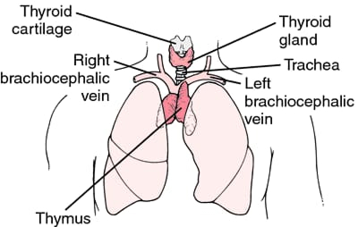
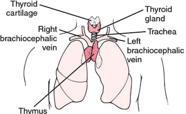
THYMUS GLAND:
It is a Bi-lobed lymphatic organ.
Thymus gland is located in Mediastinum.
Its weight is 10 to 15 gm at birth.
It reaches to its maximum size at the age of 10 to 12 years I.e 40 gm
ORGANS ASSOCIATED WITH THYMUS GLAND :
ANTERIOR: STERNUM AND UPPER 4 COASTAL CARTILAGES.
POSTERIOR : AORTIC ARCH, TRACHEА
LATERAL : LUNGS
SUPERIOR: STRUCTURES IN THE ROOT OF THE NECK
INFERIOR : HEART
STRUCTURE OF THYMUS GLAND :
The Thymus gland consists of two lobes.
Each lobe is divided into lobules.
Each lobule consists of outer cortex and central Medullary part.
These lobules consists of an irregular branching framework of epithelial cells and lymphocytes.
FUNCTION OF THYMUS GLAND:
Help in maturation of T Lymphocytes.
T Lymphocyte production occurs from thymic process of Thymus gland.
Thymosin stimulate the maturation of Thymus gland and other lymphoid organ.
This hormone is secreted by epithelial cells.
FUNCTION OF LYMPHATIC SYSTEM :
(1) DRAINING INTERSTITIAL FLUID :
Lymphatic vessels drain excess interstitial fluid to blood circulation.
(2) TRANSPORTING DIETARY LIPIDS:
Lymphatic vessels carry lipids and lipid soluble vitamins (A, D,E AND K) Absorbed by gastrointestinal tract to the Blood.
(3) PROTECTING AGAINST INVASION:
Lymphocytes aided by macrophages recognise foreign cells and microbes and respond to them in 2 basic ways:
T Lymphocyte destroy the intruder.
B Lymphocyte differentiate into plasma cells that secrete Antibodies.