UNIT 6
SPECIAL SENSES
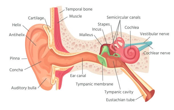
Structure of Ear :
Anatomically the Ear is divided into three regions.
External Ear
Middle Ear
Inner Ear
(1) EXTERNAL EAR :
The External Ear collects the sound waves and channels them Inward.
External Ear consists of the Auricle (Pinna) and the External Acoustic Meatus called Auditory canal.
THE AURICLE (PINNA):
Auricle or Pinna Is the flap or modified trumpet on the side of the head.
It consists of Elastic cartilage.
It helps in collection of sound.
The number of waves which enter into the Ear depends upon the size of the pinna.
EXTERNAL ACOUSTIC MEATUS (AUDITORY CANAL)
It is 2.5 centimetre long.
It is a S shaped long tube.
It lies in inward, forward and downward direction.
The wax like material is present in the canal called Cerumen.
These are modified sweat glands.
Hair are present inside the canal.
Hair and wax prevent dust or foreign material to enter in the Tympanic membrane.
The tympanic membrane is the thin layer membrane.
It separate external Ear from the Middle Ear.
It is oval shaped
(2) MIDDLE EAR :
It is the small air filled cavity.
It is present in the temporal bone.
The cavity is lined with simple squamous epithelium.
It is separated from External Ear by Ear drum.
Separated from internal Ear by Oval and round window.
The Anterior wall of the Middle Ear contains opening.
Opening leds directly into the Auditory tube.
Auditory tube is lined with bone and hyaline cartilage.
It connects the Middle Ear with the nasopharynx.
It extends downward, forward and inward.
The Auditory Tube maintains the pressure between External and internal surface of tympanic membrane, so it prevents the repture of membrane.
It also maintain pressure during yawning, high altitude and swallowing.
AUDITORY OSSICLES :
The Middle Ear contains three Auditory ossicles.
(1) MALLEUS :
It is hammer shaped bone.
The handle of the Malleus is attached to the internal surface of the Eardrum. The head of Malleus articulates with incus.
(2) INCUS:
It is the anvil shaped bone.
It is the intermediate bone.
The Malleus articulates with incus.
Incus further articulates with head of stapes.
(3) STAPES:
It is the medial stirrup shaped bone.
Its head articulates with incus.
The footplate of stapes is fit into the opening called oval window.
Below the oval window there is a round window.
(3)INNER EAR:
Inner ear is complicated.
It is the organ of Hearing and balance.
It consists of two parts.
Bony Labyrinth.
Membranous Labyrinth.
BONY LABYRINTH:
Bony labyrinth is lined with Periosteum.
It contains a fluid called Perilymph.
The bony labyrinth consists of
1 vestibule
1 cochlea
3 semicircular canals.
VESTIBULE :
It contains Utricle and Saccule.
Oval and Round window are present in its lateral wall.
COCHLEA:
It is involved in hearing.
It resembles the Snails Shell.
It is a Spiral round central Bony Column.
SEMICIRCULAR CANAL:
It is involved in balance.
These are three tubes arranged so that one is situated in each of the three planes of space.
(1) MEMBRANOUS LABYRINTH :
The fluid present in membranous labyrinth is called Endolymph. It consists of
The vestibule
Cochlea
Semicircular canals
COCHLEA:
Cochlea contains three compartments.
(1) the Scala Vestibuli
(2) the Scala Media
(3) the Scala Tympani
The cochlear duct is Triangular tube shape.
The bony cochlea is divided into upper and lower part.
Upper section is called Scala Vestibuli.
Lower section is called scala tympani.
The roof of the cochlear membrane is called Basiliar Membrane.
The hearing organ that is organ of Corti rests on Basiliar membrane.
PHYSIOLOGY OF HEARING :
The Audible Range is 20 to 20,000 hertz.
Every sound produce sound waves.
The Auricles direct sound waves into the External Auditory canal.
The sound waves strike the eardrum.
The Ear Drum vibrates.
The Ear drum is connected to malleus which also vibrates.
Then vibration passes from Malleus to incus.
Then to the Stapes.
It further pushes the oval window.
oval window generate pressure waves in the perilymph of the cochlea.
There is a wave of motion in the Endolymph.
Nerve impulses generated pass to the vestibulocochlear nerve.
Membrane of Round window vibrates.
Nerve impulses reach hearing area in the cerebrum.
EVERY SOUND PRODUCES WAVES
↓
AURICLE
↓
AUDITORY CANAL
↓
TYMPANIC MEMBRANE
↓
OSSICLES (MIDDLE EAR)
↓
OVAL WINDOW
↓
WAVES PRODUCE IN PERILYMPH
↓
MOVES TO MEMBRANOUS LABYRINTH
↓ GOES TO
CAUSE WAVES IN ENDOLYMPH
↓
CONCENTRATES THE WAVE STIMULATES THE NEURO EPITHELIAL CELL
↓ STRIKES WITH
NERVE IMPULSES THAT PASS TO VESTIBULOCOCHLEAR NERVE
↓THEN TRANSMITTED TO
CEREBRUM WHERE SOUND WAVES ARE PERCEIVED.
PHYSIOLOGY OF BALANCE :
The vestibule and semicircular canal help to maintain the balance of the body.
The equilibrium of body is of 2 types
(1) Dynamic Equilibrium:
It is the maintenance of body position in response to rotation, acceleration.
(2) Static Equilibrium:
It is the maintenance of the position of the body in relation to force of gravity.
The Semicircular canal together with the Saccule and utricle, maintains dynamic equilibrium.
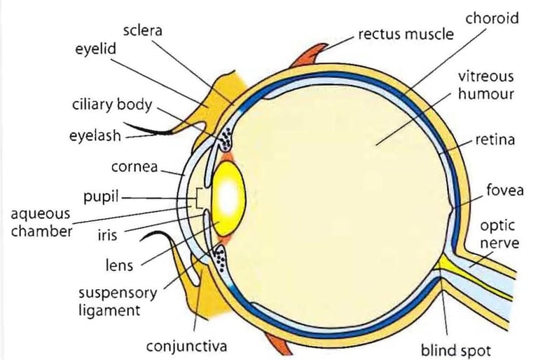
STRUCTURE OF EYE :
Eyeball is composed of three layers.
(1) Outer fibrous, Supporting layer Sclera and Cornea.
(2) Middle vascular layer Choroid, Ciliary body and Iris.
(3) Inner Nervous tissue layer Retina.
(1) Sclera and Cornea :
It is the white portion of the eye.
It is the outer fibrous layer.
The Sclera protects the delicate structures of the eye.
It helps to maintain the shape of the Eyeball.
The cornea lies over the Anterior portion of the eye.
It lies over the coloured part of the eye.
It is involved in bending light rays to focus them on the Retina.
(2)CHOROID:
It is the middle vascular layer.
It contains blood vessels.
The colour of Eye is due to choroid because it contains pigment of blue, brown, and Gray colour.
The Anterior portion is modified into three separate structures called Ciliary body, the suspension ligament and the Iris.
(3) Ciliary Body:
It Suspend the lens.
It helps in Accomodation for near vision.
Ciliary Body consists of Unstriped Ciliary muscles.
These Ciliary muscles alter the shape of the lens which help in near or far vision.
Because when these muscles contract / relax. The Thickness of lens also changes which Bend the light Rays Entering the Eye to focus them on the Retina.
(4) IRIS:
It is the coloured portion of eyeball.
Its shape is like a flattened donut.
It is located vertically between the cornea and the lens.
The Iris forms a circular curtain with an opening, in the centre called pupil.
By adjusting the size of pupil it controls the amount of light entering into the eye.
(5) LENS:
The lens is a transparent portion of the eye.
It is biconvex structure.
It is situated between anterior and posterior segments of the eye.
It is 1 centimetre in diameter.
The thickness of the lens depends upon contraction and relaxation of the Ciliary muscles.
When the object is near, the lens become thicker so as to focus the light on the retina.
(6) Retina:
It is the inner nervous tissue layer of the eyeball.
The retina contains the photo receptors, the Rods and Cones.
Each retina has about 6 million Cones, and 120 millions Rod cells.
Rod cells help to see in dim light.
Cone cell help to see in bright light that is colour vision.
The retina is Supplied by a central artery.
The small area of retina where the optic nerve leaves the eye is the optic disc Or blind spot.
It has no light sensitive cells.
Blood Supply to the Eye :
The central retinal artery and ciliary arteries Supply the blood to the eye.
These are the branches of ophthalmic artery.
The central Retinal vein helps in the venous Drainage.
Interior of the Eye:
It is divided into two cavities.
Anterior
Posterior
Aqueous humour is the fluid present in the Anterior cavity.
Vitreous humour is the fluid present in the posterior cavity.
Aqueous humour drains out of the anterior chamber at the same rate at which it enters the Posterior chamber.
In this way amount of fluid remains the same.
The normal intra-ocular pressure is 16 to 20 mm Hg.
If the intra-ocular pressure increases it leads to disease Glaucoma.
PHYSIOLOGY OF SIGHT:
The White colour is a Combination of different types of colour I.e Colours present in Rainbow.
VIBGYOR
V - VOILET
I-INDIGO
B - BLUE
G-GRAY
Y - YELLOW
O-ORANGE
R - RED
When the white light passes through the prism it bends at different angles according to its wavelength.
So it shows different colours.
Violet has shorter wavelength.
Red has greater wavelength,
The object looks white when our eyes perceive all the wavelength and black when perceived nothing that is they are all absorbed.
REFRACTION OF LIGHT RAYS:
When the light passes from one medium to another it gets bent.
For example from air to liquid, this principle is used to focus the light on the retina.
The lens helps in focusing the light.
The lens in our eye is biconvex which help in focusing the light on the retina.
PHOTORECEPTORS AND PHOTOPIGMENTS:
Both Rods and Cones contain photo-pigments.
In the Rod Cells, the Photo pigment Rhodopsin is present.
This is sensitive to light.
They are stimulated in dim light.
Photo-pigment is unable to respond to light.
Cone cells therefore function to Produce Vision in bright light. Vitamin A helps in formation of Rhodopsin, where there is bright light the Rhodopsin gets bleached and it regenerates in the presence of vitamin A.
LIGHT AND DARK ADAPΤΑΤΙΟΝ :
Our eyes are very sensitive to light.
These take some time to adjust example whenever you move from dark to light area that is from tunnel into the sunshine.
Your Visual system adjusts in seconds to brighter environment by decreasing its sensitivity this is called light adaptation.
On the other hand when you enter into a dark room example theatre your visual sensitivity increases slowly over many minutes this is called dark adaptation.
ACCESSORY ORGANS OF EYE :
(1) EYELIDS AND EYELASHES
(2)EYEBROWS
(3) LACRIMAL APPARATUS
(1) EYELIDS AND EYELASHES :
Upper and Lower eyelids shade the eyes during sleep.
It Protects the eyes from excessive light and foreign bodies.
It also lubricate the Eye l.e prevent from dryness.
Lubricating fluid is secreted by small gland.
Present at the base of Lashes.
(2) EYEBROWS:
Eyebrows serve as a cosmetic purpose.
These prevent the sweat to Enter into the Eyes.
(3) Lacrimal Apparatus:
Nourish the cornea of the Eye.
Wash away irritating materials e.g dust.
Oilness act as a lubricant.
It prevent dryness of eye.
SENSE OF SMELL :
STRUCTURE : :
It is called Olfactory sensation.
The Nasal cavity has double function l.e Sense of Smell and Passage way for Respiration.
OLFACTORY NERVE (THE FIRST CRANIAL NERVE):
One each side of the nose, Olfactory Receptors are present.
Collectively more than 40 million bundles of axons are present, forming a pathway known as olfactory Nerves.
PHYSIOLOGY OF SMELL:
Odour Producing Chemicals dissolved in the mucus surrounding the olfactory Cilia reaches a Thresh-hold level.
A Receptor potential and then an Action potential will be generated and passed to the olfactory nerves in the olfactory bulb.
The impulses passes through the olfactory tract into the olfactory centre of the brain for interpretation, integration and memory storage.
Sense of smell can create Powerful and Long lasting memories, Memories coupled to unique sensory inputs especially distinctive odours, often persist from early Childhood to death.
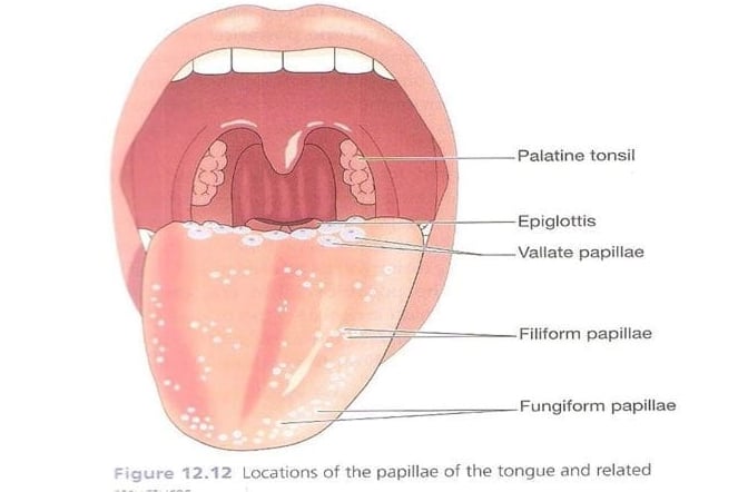
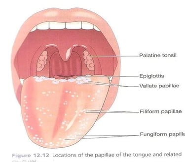
SENSE OF TASTE :
Also called Gustatory sense.
The taste buds are the sense organs.
These respond to the sense of taste.
Taste buds are chemoreceptors.
These are stimulated by chemical dissolved in the saliva.
PHYSIOLOGY OF TASTE :
Four Fundamental sensations of taste have been described as sweet, sour, bitter and salt.
Some taste stimulate taste buds in specific part of tongue like :
(1) Sweet and salty, mainly at the tip.
(2) Sour at the sides.
(3) Bitter at the back.
Sense of taste triggers salivation and the secretion of gastric juices.
It also has a protective function e.g when a foul tasting food is eaten, reflex gagging, vomiting may be induced.
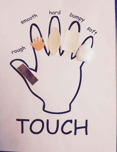
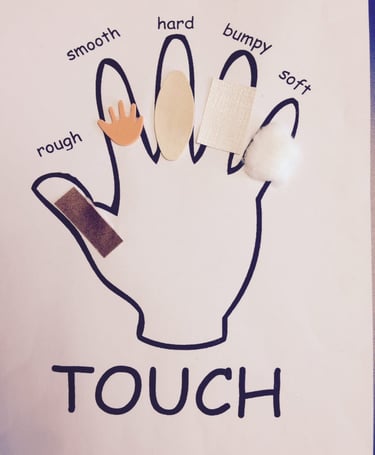
SENSE OF TOUCH:
Our Skin acts as the protective barrier b/w our internal body system and outside world.
Its ability to perceive Touch Sensations gives our Brain information about Environment around us such as Temperature, Pain, Pressure.
Without our Sense of Touch we couldn't sense when something sharp cut us and wouldn't feel, heat of sun and our Skin.
PHYSIOLOGY OF TOUCH :
Our sense of touch is controlled by huge network of nerve endings and touch receptors in the skin known as somatosensory system.
This system is responsible for all the sensations we feel cold, Hot, Smooth, Rough, Pressure, Tickle, Pain and Vibrations.
There are 4 main type of receptors :
(1) Mechano receptors
(2) Thermoreceptor
(3) Pain receptors
(4) Propriceptors